. 2008 Feb 8;283(6):3537-3549.
doi: 10.1074/jbc.M707998200. Epub 2007 Dec 10.

Direct spectroscopic study of reconstituted transcription complexes reveals that intrinsic termination is driven primarily by thermodynamic destabilization of the nucleic acid framework

Affiliations

Direct spectroscopic study of reconstituted transcription complexes reveals that intrinsic termination is driven primarily by thermodynamic destabilization of the nucleic acid framework

Kausiki Datta et al. J Biol Chem. .

Abstract

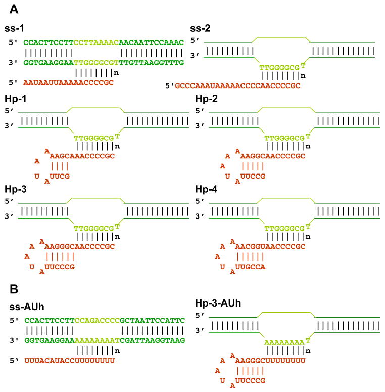
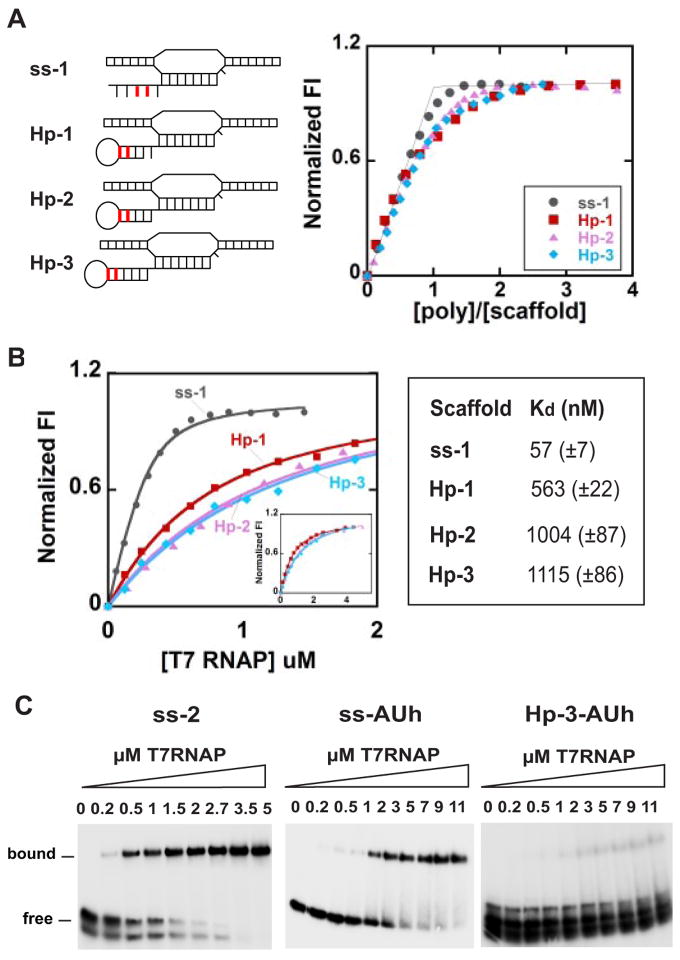
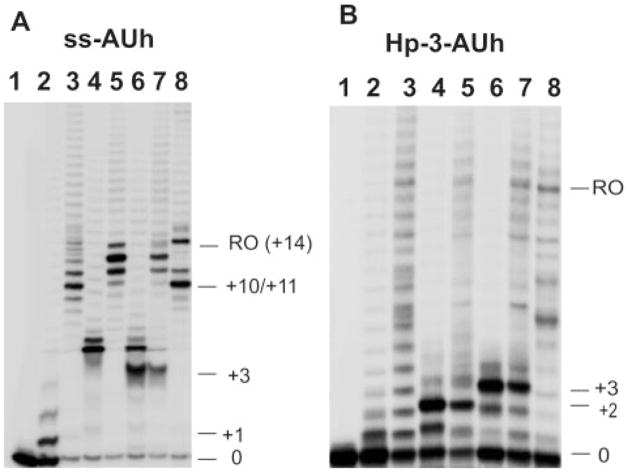
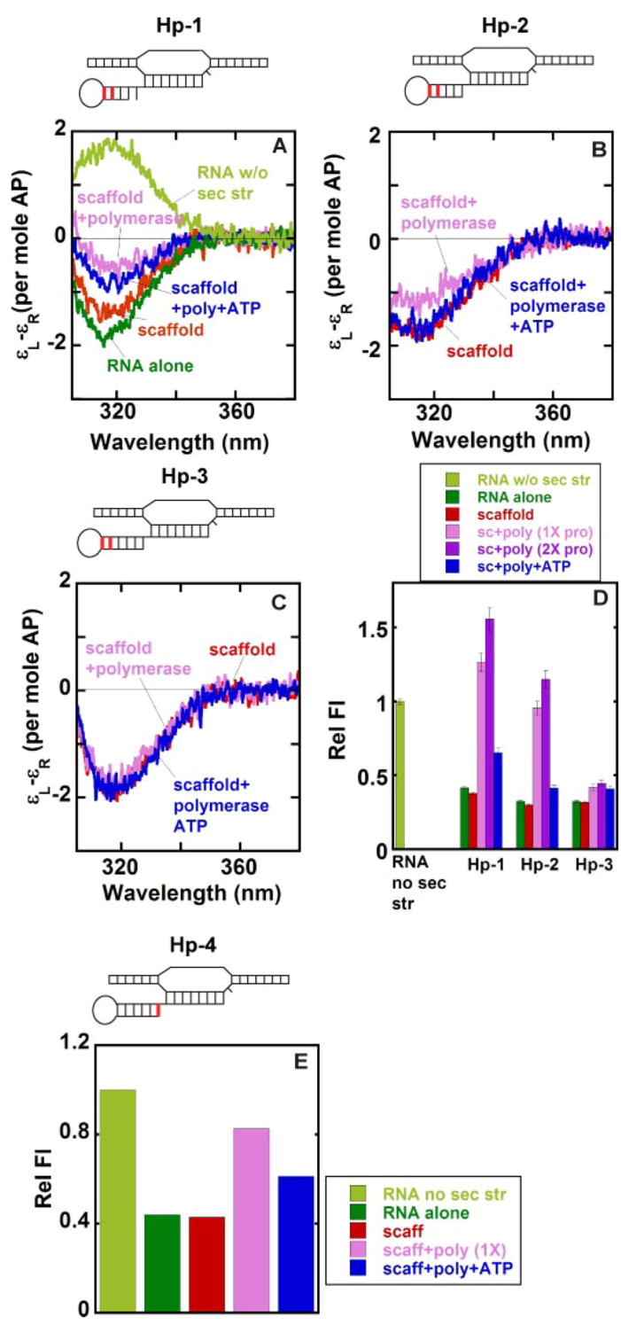
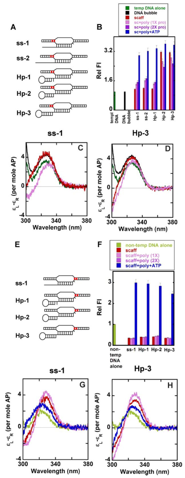
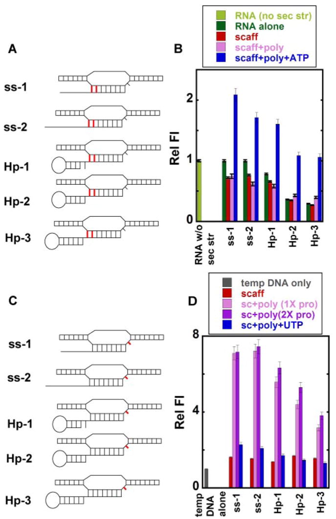
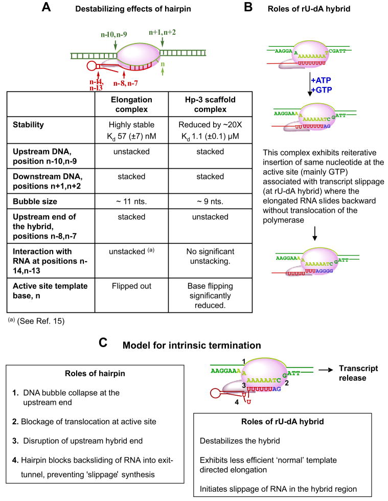
Changes in near UV circular dichroism (CD) and fluorescence spectra of site-specifically placed pairs of 2-aminopurine residues have been used to probe the roles of the RNA hairpin and the RNA-DNA hybrid in controlling intrinsic termination of transcription. Functional transcription complexes were assembled directly by mixing preformed nucleic acid scaffolds of defined sequence with T7 RNA polymerase (RNAP). Scaffolds containing RNA hairpins immediately upstream of a GC-rich hybrid formed complexes of reduced stability, whereas the same hairpins adjacent to a hybrid of rU-dA base pairs triggered complex dissociation and transcript release. 2-Aminopurine probes at the upstream ends of the hairpin stems show that the hairpins open on RNAP binding and that stem re-formation begins after one or two RNA bases on the downstream side of the stem have emerged from the RNAP exit tunnel. Hairpins directly adjacent to the RNA-DNA hybrid weaken RNAP binding, decrease elongation efficiency, and disrupt the upstream end of the hybrid as well as interfere with the movement of the template base at the RNAP active site. Probing the edges of the DNA transcription bubble demonstrates that termination hairpins prevent translocation of the RNAP, suggesting that they transiently "lock" the polymerase to the nucleic acid scaffold and, thus, hold the RNA-DNA hybrid "in frame." At intrinsic terminators the weak rU-dA hybrid and the adjacent termination hairpin combine to destabilize the elongation complex sufficiently to permit significant transcript release, whereas hairpin-dependent pausing provides time for the process to go to completion.


Figures

FIGURE 1
FIGURE 1. Nucleic acid sequences and nomenclature of the scaffold constructs used in this study
A, scaffolds with GC-rich RNA-DNA hybrid. B, scaffolds with a rU-dA hybrid. Dark green, duplex region of the DNA bubble framework; light green, non-complementary region of the DNA bubble; red, RNA transcript. n represents the active site template base.
FIGURE 2
FIGURE 2. Fluorescence and gel-shift titrations with T7 RNAP of nucleic acid scaffolds containing GC-rich and rU-dA hybrids
A, fluorescence titration curves showing the stoichiometry of binding of T7 RNAP to scaffolds containing GC-rich hybrids (the scaffold concentration was 3 μM). The 2-AP base(s) within the RNA strands of the scaffolds used is shown in red. B, fluorescence titration of the same scaffolds at 300 nM and the apparent binding affinities of T7 RNAP to these scaffolds. The inset shows the full titrations for the hairpin scaffolds. C, gel-shift assays showing binding of the ss-2, ssAUh, and Hp-3-AUh scaffolds with RNAP. Numbers at the top of the gels represent the concentration of T7 RNAP (in μM) used in the experiments shown in each lane. Scaffold concentrations were 3 μM throughout.
FIGURE 3
FIGURE 3. Transcription assays with scaffolds containing rU-dA hybrids
A and B, denaturing gels showing the elongation products formed with different NTPs with ssAUh (A) and Hp-3-AUh (B) scaffolds. Lanes 1, free scaffold; lanes 2, scaffold-RNAP complexes with 20 μM ATP; lanes 3, products in lanes 2 chased with all four NTPs; lanes 4, scaffold-RNAP complexes with 20 μM ATP and GTP; lanes 5, products in lanes 4 chased with all four NTPs; lanes 6, scaffold-RNAP complex with 20 μM ATP, GTP, and CTP; lanes 7, products in lanes 6 chased with all NTPs; lanes 8, scaffold-RNAP complexes with 1 mM concentrations of all four NTPs. The numbers on the right for all gels represent the number of nucleotide residues added to the initial RNA transcript by RNAP. RO indicates the run-off products.
FIGURE 4
FIGURE 4. Local base conformations within the stems of various free and RNAP-complexed hairpin-containing scaffolds
The 2-AP base(s) in defined positions within the RNA strand of the scaffolds is shown in red. A–C, low energy CD spectra. D and E, relative fluorescence intensities. Color coding for both the CD spectra and the fluorescence intensities: light green, control RNA without any secondary structure; dark green, hairpin-containing RNA alone; red, RNA-DNA scaffold; pink, scaffold-polymerase complex at equimo-lar concentrations; purple, scaffold-polymerase complex with twice the concentration of RNAP; blue, scaffold-RNAP complex at equimolar concentrations and RNA extended by the addition of ATP.
FIGURE 5
FIGURE 5. Local base conformations at the upstream and downstream ends of the noncomplementary DNA bubble of various free and RNAP-complexed nucleic acid scaffolds
A and E, scaffolds showing the positions of the 2-AP bases (in red) in the template and non-template strands. B and F, relative fluorescence intensities. Panels C and D and panels G and H, low energy CD spectra for ss-1 and Hp-3 scaffolds, respectively. Color coding for both the CD spectra and the fluorescence intensities: dark green, single-stranded template DNA with the 2-AP bases in the template strand (A); black, DNA bubble alone with the 2-AP bases in the template strand; light green, single-stranded non-template DNA with the 2-AP bases in the non-template strand (E); red, RNA-DNA scaffold; pink, scaffold-polymerase complex at equimolar concentrations; purple, scaffold-polymerase complex with twice the concentration of RNAP; blue, scaffold-RNAP complex at equimolar concentrations and RNA extended by the addition of ATP.
FIGURE 6
FIGURE 6. Local base conformations at the upstream end of the RNA-DNA hybrid and at the active site in free and RNAP-complexed nucleic acid scaffolds
A and C, scaffolds showing the positions of 2-AP base(s) (in red) in the RNA and in the template DNA. B and D, relative fluorescence intensities. Light green, control RNA without secondary structure (sec str); dark green, RNA alone with the 2-AP bases in the RNA strand (A); gray, single-stranded template DNA with the 2-AP base in the template DNA (C); red, RNA-DNA scaffold; pink, scaffold-polymerase complex at equimolar concentrations; purple, scaffold-polymerase complex with twice the concentration of RNAP; blue, scaffold-RNAP complex at equimolar concentrations and RNA extended by the addition of ATP (B) or UTP (D).
FIGURE 7
FIGURE 7
A, summary of the changes induced at various positions within the nucleic acid scaffold of the transcription complex by the presence of the termination hairpin. B, effects of the weak rU-dA hybrid. C, mechanistic model for the intrinsic termination process. For explanations, see boxes and “Discussion.”

References

    1. Greive SJ, von Hippel PH. Nat Rev Mol Cell Biol. 2005;6:221–232. - PubMed
    1. Arndt KM, Chamberlin MJ. J Mol Biol. 1990;213:79–108. - PubMed
    1. Wilson KS, von Hippel PH. Proc Natl Acad Sci U S A. 1995;92:8793–8797. - PMC - PubMed
    1. Platt T. Annu Rev Biochem. 1986;55:339–372. - PubMed
    1. Tahirov TH, Temiakov D, Anikin M, Patlan V, McAllister WT, Vassylyev DG, Yokoyama S. Nature. 2002;420:43–50. - PubMed

Publication types