. 2008 Feb;28(3):1068-80.
doi: 10.1128/MCB.00484-07. Epub 2007 Dec 10.

A non-tumor suppressor role for basal p19ARF in maintaining nucleolar structure and function

Affiliations

A non-tumor suppressor role for basal p19ARF in maintaining nucleolar structure and function

Anthony J Apicelli et al. Mol Cell Biol. 2008 Feb.

Abstract

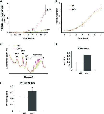
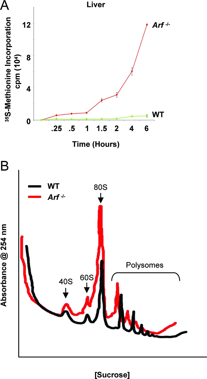
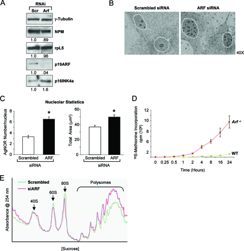
The nucleolus is the center of ribosome synthesis, with the nucleophosmin (NPM) and p19(ARF) proteins antagonizing one another to either promote or inhibit growth. However, basal NPM and ARF proteins form nucleolar complexes whose functions remain unknown. Nucleoli from Arf(-/)(-) cells displayed increased nucleolar area, suggesting that basal ARF might regulate key nucleolar functions. Concordantly, ribosome biogenesis and protein synthesis were dramatically elevated in the absence of Arf, causing these cells to exhibit tremendous gains in protein amounts and increases in cell volume. The transcription of ribosomal DNA (rDNA), the processing of nascent rRNA molecules, and the nuclear export of ribosomes were all increased in the absence of ARF. Similar results were obtained using targeted lentiviral RNA interference of ARF in wild-type MEFs. Postmitotic osteoclasts from Arf-null mice exhibited hyperactivity in vitro and in vivo, demonstrating a physiological function for basal ARF. Moreover, the knockdown of NPM blocked the increases in Arf(-/-) ribosome output and osteoclast activity, demonstrating that these gains require NPM. Thus, basal ARF proteins act as a monitor of steady-state ribosome biogenesis and growth independent of their ability to prevent unwarranted hyperproliferation.


Figures

FIG. 1.
FIG. 1.
Loss of ARF results in nucleolar morphological changes. (A) AgNOR staining of representative wild-type (WT) and Arf−/− MEFs. Increases in the number and irregularity of the AgNOR indices in the Arf−/− cells are shown. (B) Ultrastructural features of nuclei from the wild-type and Arf−/− MEFs. Arrows indicate nucleoli (×3,000) and fibrillar centers (×7,000). (C) Quantification of AgNOR indices from panel A. The left panel shows the number of AgNORs per nucleus (n = 100). The right panel shows the total nucleolar area (in μm2) per nucleus as determined by histomorphometric analysis (n = 100). *, P < 0.01.
FIG. 2.
FIG. 2.
Tissues from newborn Arf−/− mice display altered nucleolar morphology reminiscent of the in vitro findings. (A) AgNOR staining of representative sections from the intestine and liver. (B) Quantification of total AgNOR area per nucleus (n = 100). *, P < 0.01. WT, wild type.
FIG. 3.
FIG. 3.
Disruption of ARF enhances protein synthesis independent of cellular proliferation. (A) Cells were starved of methionine and cysteine for 30 min prior to the addition of a [35S]methionine label for the indicated times, followed by lysis, trichloroacetic acid precipitation of proteins, and liquid scintillation counting. (B) Equal numbers of cells (1 × 10⁵) were plated in triplicate at day 0 and then were trypsinized and counted via a hemocytometer at various time points. (C) Cycloheximide (50 μg/ml) was added for 10 min prior to lysis and ultracentrifugation of cleared lysate on 10 to 40% sucrose gradients. The graph shows an A254 of ribosome subunits over increasing sucrose density. (D) Equal-passage MEFs (1 × 10⁵) were trypsinized and analyzed by a Coulter Vi-Cell counter for cell volume. (E) Equal-passage MEFs (1 ×10⁶) were harvested and analyzed for protein content by a standard colorimetric DC assay. WT, wild type.
FIG. 4.
FIG. 4.
ARF regulates protein synthesis and ribosome biogenesis in vivo. (A) Livers were isolated from three wild-type (WT) and Arf-null littermates and briefly trypsinized. Cells (5 × 10⁶) were immediately cultured in methionine-free medium for 15 min and then incubated with [35S]methionine for the indicated times. Proteins were trichloroacetic acid precipitated, and labeled proteins were quantified by liquid scintillation counting. (B) Spleens were isolated from three wild-type and Arf-null littermates. Cells (1 × 10⁷) were immediately harvested, and cytosolic fractions were loaded onto 7 to 47% sucrose gradients for ultracentrifugation separation. Graph B shows an A254 of ribosome subunits over increasing sucrose density.
FIG. 5.
FIG. 5.
Acute depletion of p19ARF results in nucleolar, morphological, and functional changes reminiscent of the Arf−/− cells. (A) Western blotting confirmation of the p19ARF knockdown in wild-type (WT) MEFs 96 h postinfection with lentiviral shRNA constructs using antibodies recognizing γ-tubulin, NPM, rpL5, p19ARF, and p16INK4a. Expression change (n-fold) is marked under each panel. (B) AgNOR staining of representative MEFs infected with control (scrambled) or p19ARF-specific shRNA virus. (C) Quantification of AgNOR indices. Left panel shows the number of AgNORs per nucleus (n = 100). Right panel shows the total nucleolar area (in μm2) per nucleus as determined by histomorphometric analysis (n = 100). *, P < 0.01 (D) Total radioactivity incorporated after [35S]methionine pulse. Cells were starved of methionine and cysteine for 30 min prior to the addition of label for the indicated times, followed by lysis, trichloroacetic acid precipitation of proteins, and liquid scintillation counting. (E) Cycloheximide (50 μg/ml) was added for 10 min prior to lysis and ultracentrifugation of cleared lysate on 10 to 40% sucrose gradients. The graph shows an A254 of ribosome subunits over increasing sucrose density.
FIG. 6.
FIG. 6.
Loss of p19ARF has functional consequences on osteoclast biology. (A) BrdU incorporation in the wild-type (WT) and Arf−/ macrophages. (B) Representative TRAP staining of equal numbers of BMMs following 3 days of treatment with M-CSF and RANKL reveals an increase in multinucleated osteoclasts formed from the Arf−/− precursors. (C) The graph shows increases in TRAP-positive osteoclasts with greater than five nuclei derived from the Arf−/− bone marrow. *, P = 0.01. (D) TRAP solution assay of equal numbers of TRAP-positive cells. Cells from the wild-type (day 4 post-RANKL addition) or the Arf−/ (day 3 post-RANKL addition) precursors were lysed and incubated in a colorimetric assay with p-nitrophenyl phosphate, a substrate for TRAP. The graph shows an A405. *, P = 0.01. (E) Levels of serum TRAP 5b in Arf−/ compared to that in wild-type mice (P = 0.03; n = 5 mice in each group) as measured by ELISA.
FIG. 7.
FIG. 7.
ARF exerts its effects through the control of rRNA synthesis and processing. (A) Western blotting demonstrates that the Arf−/ MEFs do not have alterations in the levels of nucleolar proteins NPM and ribosomal protein L5. (B) Serial NPM immunoprecipitation. Wild-type cells were lysed and serially immunoprecipitated (five times) with mouse NPM antibodies. The final supernatant was concentrated and was included as a control for non-NPM binding proteins. (C) Total RNA was collected from equal numbers of asynchronously dividing cells, and quantitative real-time RT-PCR was performed with a primer specific to the mouse 47S transcript. (D) The wild-type (WT) and Arf−/ cells were pulsed with a [3H]uridine label for 30 min, followed by a chase with label-free medium for the indicated times. Total RNA was isolated from equal cell numbers, loaded onto formaldehyde-containing agarose gels, and transferred to membranes for fluorography. (E) Cells were labeled with [methyl-3H]methionine, followed by a chase with medium containing excess unlabeled methionine for the indicated times. Total RNA was isolated, and equal radioactive counts were loaded onto gels and transferred to membranes for fluorography. CPM, counts per minute.
FIG. 8.
FIG. 8.
Nucleocytoplasmic shuttling of newly synthesized ribosomes is enhanced in the absence of Arf. (A) Equal numbers of cells were pulsed with [methyl-3H]methionine and chased with unlabeled methionine-containing medium for the indicated times. Total RNA was isolated from nuclear (N) and cytoplasmic (C) fractions and subjected to fluorography. (B) Cytoplasmic fractions from the indicated times were also subjected to liquid scintillation counting to obtain a quantitative estimate of total cytoplasmic rRNA. Inset, scatter plot of data presented in panel B with best-fit lines to indicate the velocity of export. m = slope. WT, wild type.
FIG. 9.
FIG. 9.
Myc is not required for the enhanced rDNA transcription of the Arf-null MEFs. the Arf−/− MEFs (2 × 10⁶) transduced with siLuc control siRNAs or Myc siRNAs in the absence or presence of Myc-ER expressing retroviruses and 4-hyrodxytamoxifen were harvested and (A) immunoblotted with antibodies recognizing c-Myc or γ-tubulin. (B) RNA was isolated from the above cells and real-time PCR using 47S rRNA probes was performed in triplicate. WT, wild type.
FIG. 10.
FIG. 10.
NPM is required for ribosome gains in the absence of Arf. The Arf−/− MEFs (2 × 10⁶) infected with lentiviruses encoding scrambled or NPM shRNAs were (A) lysed and immunoblotted with antibodies recognizing NPM and γ-tubulin; (B) lysed and RNA isolated for real-time PCR using 47S rRNA probes; (C) labeled with [methyl-3H]methionine, followed by a chase with medium containing excess unlabeled methionine for the indicated times, isolation of total RNA and equal radioactive counts, loading onto gels, and transfer to membranes for fluorography; (D) fractionated into nuclear (N) and cytosolic (C) lysates and immunoblotted with lamin A/C and SOD or Northern blotted with probes recognizing the 18S rRNA; or (E) starved of methionine and cysteine for 30 min prior to the addition of label for the indicated times, followed by lysis, trichloroacetic acid precipitation of proteins, and liquid scintillation counting. *, P < 0.01.
FIG. 11.
FIG. 11.
Loss of NPM expression inhibits osteoclastogenesis in the Arf / cells. (A) Western blotting of macrophages infected with either control or lentivirus-targeted shRNA specific to NPM to confirm the gene knockdown. (B) TRAP staining of osteoclasts differentiated in vitro with RANKL and M-CSF for 6 days. (C) TRAP activity assay of equal numbers of osteoclasts from the indicated genotypes. *, P < 0.01. WT, wild type.

References

    1. Aubele, M., S. Biesterfeld, M. Derenzini, P. Hufnagl, H. Martin, D. Ofner, D. Ploton, and J. Ruschoff for the Committee on AgNOR Quantitation within the European Society of Pathology. 1994. Guidelines of AgNOR quantitation. Zentralbl. Pathol. 140107-108. - PubMed
    1. Ayrault, O., L. Andrique, D. Fauvin, B. Eymin, S. Gazzeri, and P. Seite. 2006. Human tumor suppressor p14ARF negatively regulates rRNA transcription and inhibits UBF1 transcription factor phosphorylation. Oncogene 257577-7786. - PubMed
    1. Ayrault, O., L. Andrique, C. J. Larsen, and P. Seite. 2004. Human Arf tumor suppressor specifically interacts with chromatin containing the promoter of rRNA genes. Oncogene 238097-8104. - PubMed
    1. Bertwistle, D., M. Sugimoto, and C. J. Sherr. 2004. Physical and functional interactions of the Arf tumor suppressor protein with nucleophosmin/B23. Mol. Cell. Biol. 24985-996. - PMC - PubMed
    1. Bertwistle, D., F. Zindy, C. J. Sherr, and M. F. Roussel. 2004. Monoclonal antibodies to the mouse p19(Arf) tumor suppressor protein. Hybrid. Hybridomics 23293-300. - PubMed

Publication types

LinkOut - more resources