. 2007 Dec 24;204(13):3257-69.
doi: 10.1084/jem.20070166. Epub 2007 Dec 10.

Role of host tissues for sustained humoral effects after endothelial progenitor cell transplantation into the ischemic heart

Affiliations

Role of host tissues for sustained humoral effects after endothelial progenitor cell transplantation into the ischemic heart

Hyun-Jai Cho et al. J Exp Med. .

Abstract

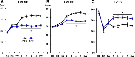
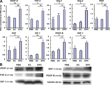
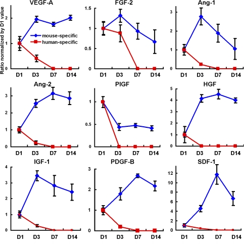
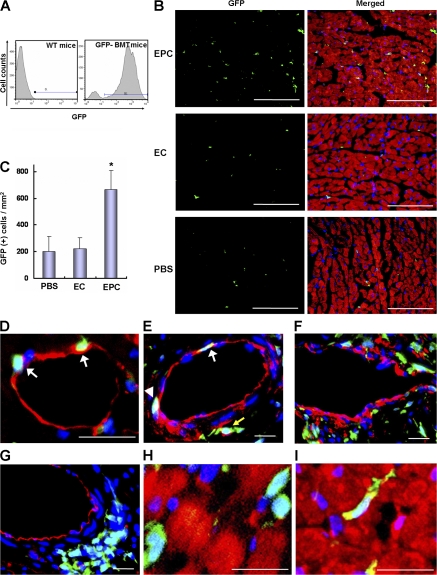
Noncellular differentiation effects have emerged as important mechanisms mediating therapeutic effects of stem or progenitor cell transplantation. Here, we investigated the expression patterns and sources of humoral factors and their regional and systemic biological effects after bone marrow (BM)-derived endothelial progenitor cell (EPC) transplantation into ischemic myocardium. Although most of the transplanted EPCs disappeared within a week, up-regulation of multiple humoral factors was sustained for longer than two weeks, which correlated well with the recovery of cardiac function. To determine the source of the humoral factors, we injected human EPCs into immunodeficient mice. Whereas the expression of human EPC (donor)-derived cytokines rapidly decreased to a nondetectable level within a week, up-regulation of mouse (recipient)-derived cytokines, including factors that could mobilize BM cells, was sustained. Histologically, we observed higher capillary density, a higher proliferation of myocardial cells, a lower cardiomyocyte apoptosis, and reduced infarct size. Furthermore, after EPC transplantation, BM-derived stem or progenitor cells were increased in the peripheral circulation and incorporated into the site of neovascularization and myocardial repair. These data indicate that myocardial EPC transplantation induces humoral effects, which are sustained by host tissues and play a crucial role in repairing myocardial injury.


Figures

Figure 1.
Figure 1.
Temporal expression patterns of multiple cytokines in peri-infarct myocardium after EPC transplantation. (A) qRT-PCR was performed to measure the level of gene expression from the samples harvested from the peri-infarct myocardium of C57BL mice at baseline (before MI, day 0), 1, 7, 14, 28, and 42 d after MI. Compared with the PBS-injected hearts, hearts transplanted with syngeneic mouse EPCs expressed higher levels of measured cytokines during the study period. Of note, cytokine levels were maintained at, or higher than, baseline levels for at least 14 d only in the EPC-transplanted hearts (n = 5 per group). *, P < 0.05; **, P < 0.01. (B and C) Immunoblots (B) and their quantification (C) showed that the protein levels of VEGF correlated well with the mRNA expression levels (n = 3 per group). *, P < 0.05; **, P < 0.01.
Figure 2.
Figure 2.
Therapeutic effects of EPC transplantation occur within the first 2 wk. Cardiac function measured by M-mode echocardiography was better in EPC-transplanted hearts compared with the control from 2 wk after MI represented by smaller LV end systolic dimension (LVESD) (A) and LV end-diastolic dimension (LVEDD) (B), and greater fractional shortening (C) (n = 7 per group). *, P < 0.05.
Figure 3.
Figure 3.
Engrafted EPCs faded away within a week after transplantation. (A) EPCs isolated from GFP-expressing mice were injected directly into the myocardium of wild-type mice. Shown are the histological sections of myocardium harvested at predetermined time points. Despite the robust initial engraftment of transplanted EPCs (green) at day 3, a majority of transplanted cells faded away within a week, and only a few transplanted cells were detected in the peri-infarct area at days 7 and 14. green, GFP; red, anti–α-sarcomeric actinin; blue, DAPI. (B) Representative figures from serial cardiac samples (days 3, 7, and 14) obtained from female mouse hearts transplanted with male mouse EPCs (wild-type). The panels show a gradual decrease of transplanted EPCs over 14 d. Red fluorescence within DAPI+ nuclei (blue) represents Y chromosome signals detected by FISH. Bars: A, 200 μm; B, 20 μm.
Figure 4.
Figure 4.
Multiple humoral factors are up-regulated after EPC transplantation. (A) qRT-PCR using tissues obtained from the peri-infarct myocardium of wild-type mice at day 14 after treatment demonstrated that various angiogenic, antiapoptotic, and chemoattractant cytokines were up-regulated in the mouse EPC-transplanted hearts compared with the PBS- or mouse EC–injected hearts. Individual values were normalized to GAPDH. Data are presented as fold difference compared with the PBS group (n = 8 per group). *, P < 0.05; **, P < 0.01. (B) Immunoblots showed that representative humoral factors were also up-regulated at the protein level. Protein expression correlated well with mRNA expression. Blots represented at least four independent experiments.
Figure 5.
Figure 5.
Host cells constitute the major role for sustained up-regulation of the humoral factors. To determine whether the up-regulated cytokines were derived from injected donor cells or recipient host cells, human EPCs were transplanted into immunocompromised nude mice, and the levels of cytokines were measured by both human- and mouse-specific primers and probes for each cytokine. The expression levels of cytokines from human EPCs (donor cells) were at their highest levels at day 1 and fell to an undetectable range within 7 d. Most of the mouse (host)-specific cytokine levels continued to rise after day 1 and were maintained at higher than the baseline levels over 14 d (n = 5 per each time point). Individual values were normalized to GAPDH and shown as fold difference to the values at day 1.
Figure 6.
Figure 6.
EPC transplantation decreases myocardial apoptosis and augments proliferation of myocardial cells. (A and B) Concomitant staining of EPC-, EC-, and PBS-treated myocardium with α-sarcomeric actinin (red) and TUNEL (green) to identify apoptotic cardiomyocytes (arrows) at day 7 after MI (A). Fewer apoptotic cells were evident in the peri-infarct area of mice receiving EPCs compared with those receiving EC or PBS (B) (n = 7 per group). *, P < 0.01. (C and D) Identification of proliferating cells by Ki-67 IHC at 14 d after MI. In myocardial sections, Ki-67+ cells are demonstrated by green fluorescence (C). Proliferating cardiomyocytes with a regular rod shape (EPC, top) and small-sized elongated shape (EPC, bottom) were more frequently observed in the EPC group. Quantification revealed that EPC-transplanted hearts had a significantly higher number of Ki-67+ myocardial cells compared with the EC- or PBS-injected hearts (n = 7 per group). *, P < 0.01 versus EC and PBS groups. Bars: A, 100 μm; C, left and middle, 100 μm; C, right, 20 μm.
Figure 7.
Figure 7.
Intramyocardial transplantation of EPCs enhances mobilization of EPCs and stem cells from BM. (A and B) EPC culture assay of peripheral blood demonstrated that the number of circulating EPCs, which were defined as adherent cells that showed the characteristics of acetylated LDL uptake (red color) and lectin binding (green color), significantly increased in the EPC group compared with other control groups at days 7 and 14 after MI. *, P < 0.01 versus EC and PBS at day 7; **, P < 0.01 versus EC and PBS at day 14. (C and D) Flow cytometry analysis showed that the number of double-positive cells for VEGFR-2 and Sca-1 epitopes was significantly increased in the EPC group at day 14. *, P < 0.01 versus EC and PBS groups. (E–G) Measurement of circulating linc-kit+Sca1+ cells by flow cytometry analysis. Hematopoietic lineage cells were selected by R2 gate from peripheral blood mononuclear cells after staining with hematopoietic lineage cocktail antibodies (E). Such Lin cells within the R2 gate were analyzed by c-kit and Sca-1 markers in EPC, EC, and PBS groups (F). Quantitative analysis showed that Linc-kit+Sca-1+ cells in peripheral circulation were significantly increased in the EPC-transplanted group compared with the control groups at day 7 (n = 5 per group). *, P < 0.05 versus EC and PBS at day 7. Bars: A, 40 μm.
Figure 8.
Figure 8.
EPC transplantation augments incorporation of BM-derived cells into ischemic hearts. (A) After lethal irradiation, the BM of wild-type mice was reconstituted with BM cells isolated from eGFP transgenic mice. 8 wk after BMT, flow cytometry analysis showed that >90% of peripheral blood mononuclear cells expressed GFP protein, suggesting successful reconstitution of BM. (B) At day 14 after induction of MI in these chimeric mice, the recruited cells from BM to the peri-infarct area were evaluated. Green, GFP; red, α-sarcomeric actinin; blue, DAPI for nuclei. (C) Quantification of GFP+ cells showed that EPC transplantation significantly augmented the incorporation of BM-derived cells into the peri-infarct area (n = 5 per group). *, P < 0.01 versus EC and PBS at day 14. (D–G) IHC with Isolectin B4 (D, red) or CD31 (E–G, red) revealed the localization of BM-derived GFP+ cells within and around the vasculature in the peri-infarct area at day 14 after MI. White arrows indicate GFP+ cells, which are both incorporated into vasculature and colocalized with ECs, suggestive of transdifferentiation. A white arrowhead points to the pericytic localization of GFP+ cells, and a yellow arrow illustrates perivascular localization of GFP+ cells. The majority of GFP+ cells were located in the pericytic (F) and perivascular area (G). (H) IHC with α-sarcomeric actinin (red) revealed that the majority of BM-derived cells did not exhibit a cardiomyocyte phenotype. (I) Rarely, a few GFP+ cells (arrow) showed immature cardiomyocyte morphology. Bars: B, 100 μm; D–I, 20 μm.
Figure 9.
Figure 9.
EPC transplantation decreases infarct size and increases capillary density. (A) Representative picrosirius red staining of hearts 14 d after MI. The red color represents fibrosis. (B) EPC transplantation significantly reduced percent circumferential fibrosis (n = 7 per group). *, P < 0.05. (C) Isolectin B4 staining in the peri-infarct area (red color) at day 14. (D) EPC transplantation significantly increased capillary density (n = 7 per group). *, P < 0.01 versus EC and PBS groups. Bars: C, 100 μm.

References

    1. WHO. World Health Report. www.who.int/whr/
    1. Orlic, D., J. Kajstura, S. Chimenti, F. Limana, I. Jakoniuk, F. Quaini, B. Nadal-Ginard, D.M. Bodine, A. Leri, and P. Anversa. 2001. Mobilized bone marrow cells repair the infarcted heart, improving function and survival. Proc. Natl. Acad. Sci. USA. 98:10344–10349. - PMC - PubMed
    1. Orlic, D., J. Kajstura, S. Chimenti, I. Jakoniuk, S.M. Anderson, B. Li, J. Pickel, R. McKay, B. Nadal-Ginard, D.M. Bodine, et al. 2001. Bone marrow cells regenerate infarcted myocardium. Nature. 410:701–705. - PubMed
    1. Yoon, Y.S., A. Wecker, L. Heyd, J.S. Park, T. Tkebuchava, K. Kusano, A. Hanley, H. Scadova, G. Qin, D.H. Cha, et al. 2005. Clonally expanded novel multipotent stem cells from human bone marrow regenerate myocardium after myocardial infarction. J. Clin. Invest. 115:326–338. - PMC - PubMed
    1. Kocher, A.A., M.D. Schuster, M.J. Szabolcs, S. Takuma, D. Burkhoff, J. Wang, S. Homma, N.M. Edwards, and S. Itescu. 2001. Neovascularization of ischemic myocardium by human bone-marrow-derived angioblasts prevents cardiomyocyte apoptosis, reduces remodeling and improves cardiac function. Nat. Med. 7:430–436. - PubMed

Publication types