. 2007 Dec 24;204(13):3085-93.
doi: 10.1084/jem.20071003. Epub 2007 Dec 10.

A human postnatal lymphoid progenitor capable of circulating and seeding the thymus

Affiliations

A human postnatal lymphoid progenitor capable of circulating and seeding the thymus

Emmanuelle M Six et al. J Exp Med. .

Abstract

Identification of a thymus-seeding progenitor originating from human bone marrow (BM) constitutes a key milestone in understanding the mechanisms of T cell development and provides new potential for correcting T cell deficiencies. We report the characterization of a novel lymphoid-restricted subset, which is part of the lineage-negative CD34(+)CD10(+) progenitor population and which is distinct from B cell-committed precursors (in view of the absence of CD24 expression). We demonstrate that these Lin(-)CD34(+)CD10(+)CD24(-) progenitors have a very low myeloid potential but can generate B, T, and natural killer lymphocytes and coexpress recombination activating gene 1, terminal deoxynucleotide transferase, PAX5, interleukin 7 receptor alpha, and CD3epsilon. These progenitors are present in the cord blood and in the BM but can also be found in the blood throughout life. Moreover, they belong to the most immature thymocyte population. Collectively, these findings unravel the existence of a postnatal lymphoid-polarized population that is capable of migrating from the BM to the thymus.


Figures
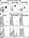
Figure 1.
Figure 1.
Flow cytometry analysis of cord blood and postnatal BM LinCD34+ progenitors. Mononuclear cells from postnatal BM and CD34+-enriched cord blood were stained with a combination of lineage (Lin) markers and anti-CD34, anti-CD10, and anti-CD24 antibodies. (A) Representative analysis of CD10 and CD24 expression of gated LinCD34+ cells from cord blood and BM of young (<10 yr of age) and older (10–60 yr of age) donors. (B) CCR9 and CCR7 expression of gated LinCD34+CD10, LinCD34+CD10+CD24, and LinCD34+CD10+CD24+ cells (shaded histogram). The dotted line indicates control isotypes. Numbers in A and B represent percentages of cells.
Figure 2.
Figure 2.
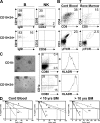
Differentiation potential of CD10+CD24 and CD10CD24+ progenitors. (A) Flow cytometry analysis of 3-wk cultures on MS5 stromal cells in B or NK cell differentiation assays of CD10+CD24 and CD10+CD24+ progenitors sorted from <10-yr-old BM. The data shown represent six independent experiments performed on cord blood (n = 2) and <10-yr-old (n = 2) and >10-yr-old (n = 2) BM samples. (B) CD4, CD8, CD3, and γδTCR expression analysis of 5-wk cultures on OP9-hDelta1 stromal cells of CD10+CD24 progenitors sorted from cord blood or pediatric BM. The data shown represent five independent experiments performed on cord blood (n = 2) and <10-yr-old (n = 2) and >10-yr-old (n = 1) BM samples. After 5 wk, we recovered an average of 6,000 and 200 T cells (CD4+CD8+ and γδ T cells) from 1,000 CD10+CD24 progenitors sorted from cord blood and BM samples, respectively. (C) LinCD34+CD10+ progenitors exhibit a DC potential. (left) A representative 10-d culture in DC differentiation conditions from cord blood–sorted CD10 and CD10+CD24 progenitors is shown. Bar, 10 μm. In the same culture conditions, CD10+CD24+ cells did not survive. (right) Flow cytometry analysis of 10-d cultures of CD10 and CD10+CD24 progenitors. HLA-DR expression is analyzed on CD1a+-gated cells. Numbers in A–C represent percentages of cells. (D) Limiting dilution assay analysis of CD10+CD24 progenitors sorted from cord blood (n = 2) and <10-yr-old (n = 1) and >10-yr-old (n = 1) BM. After 4–5 wk of culture in 96-well plates, means of 20,000, 3,250, and 560 viable cells were recovered from 100 CD10+CD24 progenitors sorted from cord blood, pediatric, and adult BM samples, respectively.
Figure 3.
Figure 3.
Flow cytometry analysis and differentiation potential of LinCD34+ cells in adult blood and postnatal thymus. (A) CD34+ enriched cells isolated from the blood of an adult healthy donor were stained with a combination of lineage (Lin) markers and anti-CD34, anti-CD7, and anti-CD10 antibodies. Data are representative of 5 pediatric and 11 adult blood samples. (B) CD10+ and CD10 progenitors recovered from adult blood were plated in B/NK (left) or T (right) cell differentiation conditions. The culture was analyzed using flow cytometry. The results of one out of four experiments are shown. (C) CD34+-enriched cells from postnatal thymus were stained with a combination of lineage (Lin) markers and anti-CD34, anti-CD7, anti-CD1a, and anti-CD10 antibodies. (left) Analysis of Lin and CD34 expression in CD34+-enriched thymocytes. (right) CD10/CD7 expression on gated LinCD34+ cells is shown. CD1a expression is analyzed on LinCD34+CD10+CD7 and CD7+ cells (histogram, bottom). Data are representative of 25 analyzed thymuses. (D) The lymphoid potential of LinCD34+CD10+ thymic progenitors. (left) Up to 8,000 LinCD34+CD10+CD7 thymic progenitors were plated in B/NK cell differentiation conditions and analyzed after 3 wk. (right) Flow cytometry analysis of a representative 5-wk culture on OP9-hDelta1 stromal cells from postnatal thymus-sorted LinCD34+CD10+CD7 cells. After 5 wk, we recovered an average of 4,000 T cells (CD4+CD8+ and γδ T cells) from 500 CD10+CD7 progenitors sorted from thymus samples. Numbers in A–D represent percentages of cells.
Figure 4.
Figure 4.
Gene expression profile of CD10, CD10+CD24, and CD10+CD24+ progenitors. (A) BM or (B) cord blood LinCD34+CD10, LinCD34+CD10+CD24, and LinCD34+CD10+CD24+ populations and (C) thymic LinCD34+CD10+CD24CD7 cells were sorted directly into PCR tubes at 10 cells per well. RT followed by a quadruplex or triplex amplification PCR assay was performed. Each well was duplicated to perform a second qualitative PCR, and seven or eight representative wells are presented for each population. DNA fragment sizes are indicated. −, negative control.

References

    1. Donskoy, E., and I. Goldschneider. 1992. Thymocytopoiesis is maintained by blood-borne precursors throughout postnatal life. A study in parabiotic mice. J. Immunol. 148:1604–1612. - PubMed
    1. Kondo, M., I.L. Weissman, and K. Akashi. 1997. Identification of clonogenic common lymphoid progenitors in mouse bone marrow. Cell. 91:661–672. - PubMed
    1. Kouro, T., K.L. Medina, K. Oritani, and P.W. Kincade. 2001. Characteristics of early murine B-lymphocyte precursors and their direct sensitivity to negative regulators. Blood. 97:2708–2715. - PubMed
    1. Bhandoola, A., and A. Sambandam. 2006. From stem cell to T cell: one route or many? Nat. Rev. Immunol. 6:117–126. - PubMed
    1. Krueger, A., A.I. Garbe, and H. von Boehmer. 2006. Phenotypic plasticity of T cell progenitors upon exposure to Notch ligands. J. Exp. Med. 203:1977–1984. - PMC - PubMed

Publication types