

Hydatid disease involving some rare locations in the body: a pictorial essay
- PMID: 18071284
- PMCID: PMC2627456
- DOI: 10.3348/kjr.2007.8.6.531
Hydatid disease involving some rare locations in the body: a pictorial essay
Abstract
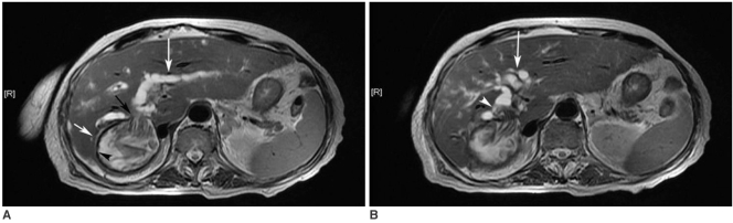
Hydatid disease (HD) is an endemic illness in many countries, and it poses an important public health problem that's influenced by peoples' socioeconomic status and migration that spreads this disease. Although rare, it may occur in any organ or tissue. The most common site is the liver (59-75%), followed in frequency by lung (27%), kidney (3%), bone (1-4%) and brain (1-2%). Other sites such as the heart, spleen, pancreas and muscles are very rarely affected. Unusual sites for this disease can cause diagnostic problems. This pictorial essay illustrates various radiological findings of HD in the liver, spleen, kidney, pancreas, peritoneal cavity, omentum, adrenal, ovary, lung, mediastinum and retroperitoneum. Familiarity with the imaging findings of HD may be helpful in making an accurate diagnosis and preventing potential complications.
Figures


References
-
- Lewall DB. Hydatid disease: biology, pathology, imaging and classification. Clin Radiol. 1998;53:863–874. - PubMed
-
- Polat P, Kantarci M, Alper F, Suma S, Koruyucu MB, Okur A. Hydatid disease from head to toe. Radiographics. 2003;23:475–494. - PubMed
-
- Pedrosa I, Saiz A, Arrazola J, Ferreiros J, Pedrosa CS. Hydatid disease: radiologic and pathologic features and complications. Radiographics. 2000;20:795–817. - PubMed
-
- Czermak BV, Unsinn KM, Gotwald T, Niehoff AA, Freund MC, Waldenberger P, et al. Echinococcus granulosus revisited: radiologic patterns seen in pediatric and adult patients. AJR Am J Roentgenol. 2001;177:1051–1056. - PubMed
-
- Beggs I. The radiology of hydatid disease. AJR Am J Roentgenol. 1985;145:639–648. - PubMed
MeSH terms
Substances
LinkOut - more resources
Full Text Sources
