. 2008 Jan;16(1):146-53.
doi: 10.1038/sj.mt.6300343. Epub 2007 Dec 11.

Exponential enhancement of oncolytic vesicular stomatitis virus potency by vector-mediated suppression of inflammatory responses in vivo

Affiliations

Exponential enhancement of oncolytic vesicular stomatitis virus potency by vector-mediated suppression of inflammatory responses in vivo

Jennifer Altomonte et al. Mol Ther. 2008 Jan.

Abstract

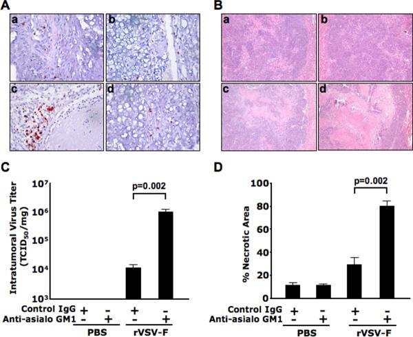
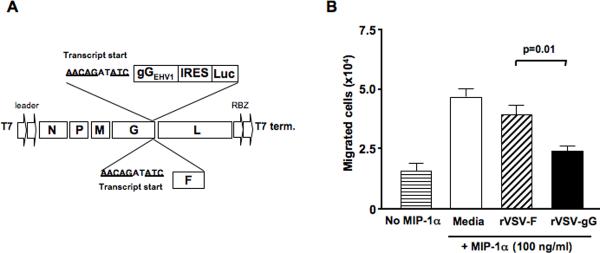
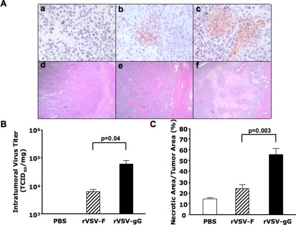
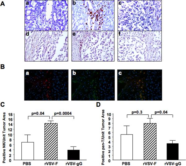
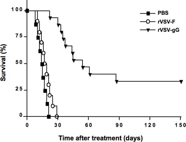
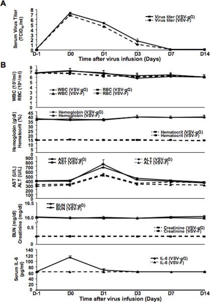
Oncolytic virotherapy is a promising strategy for treatment of malignancy, although its effectiveness is hampered by host antiviral inflammatory responses. The efficacy of treatment of oncolytic vesicular stomatitis virus (VSV) in rats bearing multifocal hepatocellular carcinoma (HCC) can be substantially elevated by antibody-mediated depletion of natural killer (NK) cells. In order to test the hypothesis that the oncotyic potency of VSV can be exponentially elevated by evasion of inflammatory responses in vivo, we constructed a recombinant VSV vector expressing equine herpes virus-1 glycoprotein G, which is a broad-spectrum viral chemokine binding protein (rVSV-gG). Infusion of rVSV-gG via the hepatic artery into immune-competent rats bearing syngeneic and multifocal HCC in their livers, resulted in a reduction of NK and NKT cells in the tumors and a 1-log enhancement in intratumoral virus titer in comparison with a reference rVSV vector. The treatment led to increased tumor necrosis and substantially prolonged animal survival without toxicities. These results indicate that rVSV-gG has the potential to be developed as an effective and safe oncolytic agent to treat patients with advanced HCC. Furthermore, the novel concept that oncolytic potency can be substantially enhanced by vector-mediated suppression of host antiviral inflammatory responses could have general applicability in the field of oncolytic virotherapy for cancer.


Figures

Figure 1
Figure 1. Logarithmic elevation of intratumoral rVSV replication and enhanced tumor necrosis by antibody-mediated depletion of NK cells in tumor-bearing rats
Buffalo rats harboring multi-focal HCC lesions in the liver were intravenously injected with polyclonal antibodies anti-asialoGM1 (Wako, Richmond, VA) or control rabbit IgG at 1mg/200μl/rat at one day before rVSV-F infusion through the hepatic artery. A single injection of rVSV-F at 1.3×10⁷ pfu/ml/rat or PBS was performed on the following day. The antibody injections were repeated via at one day after rVSV-F or PBS infusion (N=3 for each group). The treated animals were sacrificed 3 days after virus administration. Panel A shows the representative sections after immunohistochemical staining for NK cells in the four treatment groups (frame a, PBS with control rabbit IgG; frame b, PBS with anti-asialoGM1; frame c, rVSV-F with control rabbit IgG and frame d, rVSV-F with anti-asialoGM1). Panel B shows the representative sections after H&E staining in the same treatment groups as described in panel A above. Panel C shows viral titers from tumor lysates, expressed in TCID50 per mg of tumor tissue. Viral titers following treatment with rVSV-F + control Ig versus rVSV-F + Anti-asialoGM1 were statistically significant by unpaired T-test analysis (p=0.002). Panel D shows percentages of necrotic areas within tumors, as quantified by morphometric analysis of H&E- stained tumor sections. Percentages of necrosis in tumors from animals treated with rVSV-F + control IgG were compared with those treated with rVSV-F + Anti-asialoGM1 by unpaired T-test (p=0.002).
Figure 2
Figure 2. Molecular construction, rescue and functional characterization of rVSV-gG in vitro
Panel A, a schematic representation of rVSV-F and rVSV-gG. The full-length pVSV plasmid containing five transcription units, and a bicistronic construct containing the Equine Herpesvirus 1 gG (gGEHV1) and firefly luciferase (Luc), with an intervening IRES from EMCV, are shown. The transgenes are preceded by a VSV transcription termination signal, an intergenic region and a transcription start signal, and are inserted into the 3'-untranslated region of the VSVG gene. Panel B, inhibition of NK cell migration in response to MIP-1α by conditioned media from rVSV-gG infected rat HCC cells. The NK cell migration assays were performed using 24-well transwell plates. The migration of rat NK cells from the upper chamber to the lower chamber in response to 100ng/ml of MIP-1α was monitored in the presence of ultra-filtered and UV-irradiated supernatants from 105 HCC cells infected with rVSV-gG or rVSV-F. The number of NK cells in the lower chamber was counted after an incubation period of 4 hours at 37 degrees. Data presented are the mean values of four independent experiments and the results were analyzed statistically by two-sided student t test.
Figure 3
Figure 3. Intratumoral virus replication and tumor response in rats treated with rVSV-gG versus rVSV-F
Multi-focal HCC-bearing Buffalo rats were injected with PBS (n=3), rVSV-F (n=4) or rVSV-gG (n=4) at 1.3×10⁷ pfu/ml/rat and sacrificed 3 days post-virus administration via hepatic artery. Panel A, tumor sections were stained with a monoclonal anti-VSVG antibody (frames a–c) or with H&E (frames d–f). Representative sections from rats treated with PBS (frames a and d), rVSV-F (frames b and e) and rVSV-gG (frames c and f) are shown (magnification=40×). Panel B, virus titers in tumor extracts on BHK-21 cells. Viral titers are expressed as TCID50/mg tissue (mean + standard deviation), and the results were analyzed statistically by two-sided student t test (p=0.04). Panel C, tumor response as quantified by morphometric analysis using the ImagePro software. Data were shown as mean + standard deviation, and the results were analyzed statistically by two-sided student t test (p=0.003).
Figure 4
Figure 4. Immunohistochemical and immunofluorescent staining of NK and T cells in tumors of rats treated with rVSV-F versus rVSV-gG
Panel A, representative immunohistochemically stained sections from tumors and surrounding tissues. Tumor-bearing rats were infused with PBS (frames a and d), rVSV-F (frames b and e) or rVSV-gG (frames c and f) at 1.3×10⁷ pfu/ml/rat. Samples were obtained from rats at day 3 after virus infusion into the hepatic artery. Sections were stained with mouse monoclonal anti-NKR-P1A (frames a–c) or monoclonal anti-OX-52 (frames d–f) (magnification=40×). Panel B, representative immunoflurescent sections of rVSV-F infected rat HCC tumors. Frozen sections were fixed with cold acetone and blocked with 4% goat serum, followed by staining with R-PE-conjugated mouse anti-rat CD3 monoclonal antibody (frame a), FITC-conjugated mouse anti-rat NKR-P1A (frame b), and a merged picture (frames c) (magnification=40×). Panels C and D, semi-quantification of NK and T cells in the lesions after PBS, rVSV-F and rVSV-gG treatment, respectively, as quantified by morphometric analysis using the ImagePro software. Immune cell index was calculated as ratio of positive cell to unit tumor area (10,000 pixel as one unit tumor area), and the results were analyzed statistically by two-sided student t test.
Figure 5
Figure 5. Kaplan-Meier survival curve of multi-focal HCC-bearing rats after rVSV-F versus rVSV-gG treatment
HCC-bearing rats were given hepatic arterial infusion of PBS (n=8), rVSV-F (n=10) or rVSV-gG (n=15) at 1.3×10⁷ pfu/ml/rat. Survival was monitored daily and the results were analyzed statistically by log rank test (p=0.00001).
Figure 6
Figure 6. Toxicology studies of tumor-bearing rats after rVSVgG and rVSV-F administration
Panel A, kinetic profile of infectious virus yields in sera of tumor-bearing rats after infusion of rVSV-gG and rVSV-F. Virus titers (TCID50/ml) in sera obtained from tumor-bearing animals at one day before (D-1), and at 30 minutes, 1, 3, 7 and 14 days after (D0, D1, D3, D7 and D14, respectively), virus infusion into hepatice artery are shown (mena ± standard deviation; n=4–5/time point). A peak of virus titer was observed at D0 for both vectors, which declined logarithmically to undetactable levels at D7 and thereafter. Panel B, determination of CBC and serum chemistries in tumor-bearing rats after hepatic arterial infusion of rVSV-gG and rVSV-F. Blood samples were collected from the same sets of animals as in Panel A at the indicated time points. WBC, RBC, hemoglobin, hematocrit, BUN and creatinine levels in the animals remained within their respective normal ranges at all time points after administration of either vector. Serum IL-6 levels in either vector treated rats were also below its detection limit (<62.5 pg/ml) at all time points, except at D0 (~120 pg/ml) in the rVSV-gG treated rats, although the peak was more than two orders of magnitude below that considered to be toxic.

References

    1. Parkin DM, Sthernsward J, Muir CS. Estimates of the worldwide frequency of twelve major cancers. Bull World Health Organ. 1984;62:163–182. - PMC - PubMed
    1. Murray CJ, L AD. Evidence-based health policy - lessons from the Global Burden of Disease Study. Science. 1996;274:740–743. - PubMed
    1. Parkin DM, Bray F, Ferlay J, Pisani P. Estimating the world cancer burden: Globocan 2000. Int J Cancer. 2001;94:153–156. - PubMed
    1. El-Serag HB, M AC. Rising incidence of hepatocellular carcinoma in the United States. N Engl J Med. 1999;340:745–750. - PubMed
    1. Dyer Z, Peltekian K, van Zanten SV. The changing epidemiology of hepatocellular carcinoma in Canada. Aliment Pharmacol Ther. 2005;22:17–22. - PubMed

Publication types

MeSH terms

Substances

LinkOut - more resources