. 2008;25(3):201-11.
doi: 10.1007/s10585-007-9133-3. Epub 2007 Dec 11.

Inhibition of the CXCR4/CXCL12 chemokine pathway reduces the development of murine pulmonary metastases

Affiliations

Inhibition of the CXCR4/CXCL12 chemokine pathway reduces the development of murine pulmonary metastases

Su Young Kim et al. Clin Exp Metastasis. 2008.

Abstract

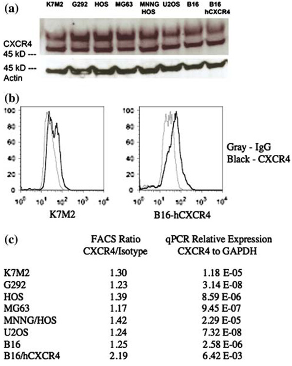
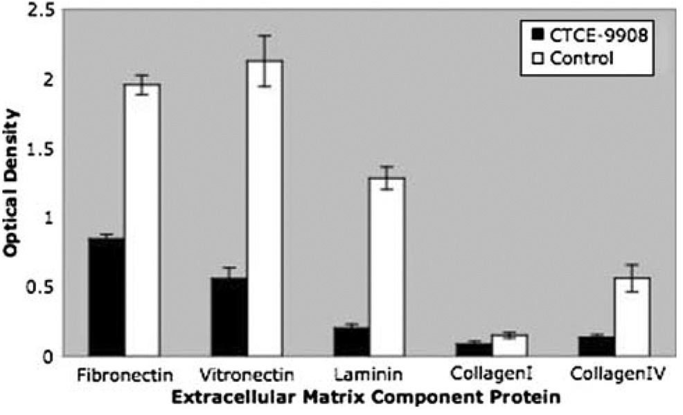
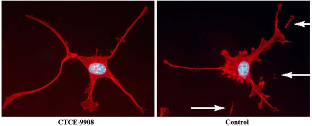
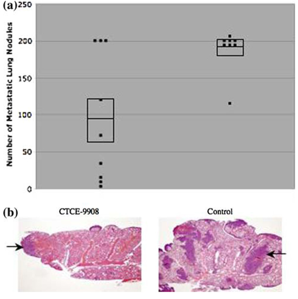
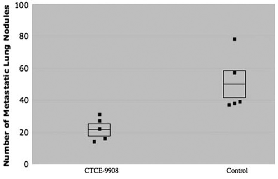
Metastasis continues to be the leading cause of mortality for patients with cancer. High expression of the chemokine receptor CXCR4 correlates with poor prognosis in many cancers, including osteosarcoma and melanoma. CXCL12, the ligand for CXCR4, is expressed at high levels in the lung and lymph node, which are the primary sites to which these tumors metastasize respectively. These findings suggest that therapy aimed at disruption of this specific receptor/ligand complex may lead to a decrease in metastases. CTCE-9908, a small peptide CXCR4 antagonist was utilized in two murine metastasis models to test this hypothesis. Treatment of osteosarcoma cells in vitro with CTCE-9908 led to the following changes: decreased adhesion, decreased migration, decreased invasion, and decreased growth rate. Following tail vein injection of osteosarcoma cells, mice that were treated with CTCE-9908 had a 50% reduction in the number of gross metastatic lung nodules and a marked decrease in micro-metastatic disease. Similar findings were observed following injection of melanoma cells and treatment with CTCE-9908. However, these results could only be consistently reproduced when the cells were pre-treated with the inhibitor. A novel ex vivo luciferase assay showed decreased numbers of cells in the lung immediately after injection into mice, when treated with CTCE-9908, suggesting the importance of interactions between the receptor and the ligand. Our findings show that inhibition of the CXCR4/CXCL12 pathway decreases metastatic disease in two murine tumor models and expands on previous reports to describe potential mechanisms of action.


Figures

Fig. 1
Fig. 1. Osteosarcoma cell lines express low levels of CXCR4
(a) Western blot analysis shows that all osteosarcoma cell lines express a similar level of CXCR4, which is known to be low from the literature. Actin is shown as a loading control. (b) FACS analysis demonstrates cell surface expression of CXCR4 in K7M2 osteosarcoma cells and B 16 melanoma cells, the latter was transfected with a human CXCR4 plasmid. (c) Quantitation of CXCR4 cell surface expression over that of IgG isotype control after FACS analysis and quantitation of CXCR4 mRNA levels relative to that of GAPDH control after quantitative RT-PCR analysis
Fig. 2
Fig. 2. CTCE-9908 decreases the growth rate of K7M2 cells in vitro
(a) K7M2 cells treated with CTCE-9908 have a decrease in growth rate as determined by optical density, shown on the Y-axis, following treatment with MTT. Control cells have a doubling time of 20 h whereas CTCE-9908 treated cells have a doubling time of 27 h. (b) Microscopic visualization of treated and untreated cells at 72 h show no phenotypic differences. This finding, and the fact that cells began their growth divergence after 25 h, suggests that the decrease in proliferative capability is not due to cytotoxicity of the compound
Fig. 3
Fig. 3. CTCE-9908 decreases adhesion to extracellular matrix proteins
K7M2 cells treated with CTCE-9908 overnight have decreased adhesion to microtiters plates coated with various extracellular matrix component proteins compared to untreated control cells. The Y axis shows optical density following crystal violet staining of cells that had remained adherent to the plate. The percentage decreases were as follows: fibronectin (56%), vitronectin (73%), laminin (84%), collagen type I (41%) and collagen type IV (76%)
Fig. 4
Fig. 4. CTCE-9908 treatment suggests decreased adhesion to the lung surface ex vivo
K7M3-luciferase cells were injected into the tail vein of Balb/C mice. Following the intraperitoneal injection of luciferin, the lungs were harvested, inflated and soaked in PBS containing luciferin. Luminescent intensity was quantiated using the Xenogen IVIS system. (a) Bright-field and overlapping luminescent intensity of lungs, 6 h after injection of CTCE-9908 treated K7M3-luciferase cells, or PBS treated K7M3-luciferase cells. (b) Quantitation of luminescence at either 30 min or 6 h after injection of CTCE-9908 treated or untreated cells. Cells that were treated with CTCE-9908 had a 2.5-fold decrease in luminescent intensity at 30 min (p = 0.03) and a 3-fold decrease in luminescent intensity at 6 h (p < 0.01)
Fig. 5
Fig. 5. CTCE-9908 decreases the migratory phenotype
The photographs show CTCE-9908 treated and untreated K7M2 cells following labeling with the F-actin specific stain Texas Red-phalloidin and the nuclear stain DAPI. Untreated cells (right panel) have numerous pseudomembranous detachments (shown with arrows). These are thought to be remnants of the cell that remain attached to the glass slide as the cell moves, consistent with the highly migratory nature of K7M2 cells. CTCE-9908 treated cells (left panel) do not show this phenotype, suggesting decreased motility. There were no morphologically detectable differences in the F-actin cytoskeleton between treated and untreated cells
Fig. 6
Fig. 6. CTCE-9908 decreases migration and invasion in the presence of CXCL12
(a) K7M2 cells treated with CTCE-9908 have a decrease in their ability to migrate through an 8-micron membrane. The Y axis represents the aggregate fluorescent intensity of calcein stained cells that were able to penetrate the membrane to the lower chamber, which contained CXCL12 as the chemo-attractant. Cells that were treated with CTCE-9908 had a 23% decrease (p < 0.01) in their ability to migrate through the membrane. (b) An even greater decrease was detected when cells were added to a matrigel coated membrane. Cells treated with CTCE-9908 had a 45% decrease (p < 0.01) in their ability to invade through the matrigel layer
Fig. 7
Fig. 7. CTCE-9908 decreases the number of lung nodules and micrometastatic disease in osteosarcoma
(a) Mice that were treated with CTCE-9908 had a 50% decrease in the number of metastatic lung nodules, shown on the Y-axis, compared to control mice (CTCE-9908 average 94.8 ± 86.7 vs. control average 189.4 ± 30.1, p = 0.01). Each dot represents the number of nodules for one mouse. The middle line in the box represents the average number of nodules while the top and bottom lines of the box represent the standard error of the mean. (b) CTCE-9908 treated mice had a marked decrease in micrometastatic disease (dark purple/blue areas with H + E staining, two of which are shown with arrows). The samples in panel B are from a repeat experiment and not from the mice depicted in panel A
Fig. 8
Fig. 8. CTCE-9908 decreases the number of lung nodules in mice injected with B16 melanoma cells
Mice were injected intravenously with B16 melanoma cells transduced to overexpress CXCR4. Mice treated with CTCE-9908 had a 56% reduction in the number of metastatic lung nodules (CTCE-9908 average 22.0 ± 7.2 vs. control average 49.8 ± 17.8, p = 0.02)

References

    1. Murdoch C. CXCR4: chemokine receptor extraordinaire. Immunol Rev. 2000;177:175–184. - PubMed
    1. Balkwill F. Cancer and the chemokine network. Nat Rev Cancer. 2004;4:540–550. - PubMed
    1. Luster AD. Chemokines—chemotactic cytokines that mediate inflammation. N Engl J Med. 1998;338:436–445. - PubMed
    1. Feng Y, Broder CC, Kennedy PE, et al. HIV-1 entry cofactor: functional cDNA cloning of a seven-transmembrane, G protein-coupled receptor. Science. 1996;272:872–877. - PubMed
    1. Muller A, Homey B, Soto H, et al. Involvement of chemokine receptors in breast cancer metastasis. Nature. 2001;410:50–56. - PubMed

MeSH terms