. 2008 Jan 9;130(1):93-102.
doi: 10.1021/ja071516m. Epub 2007 Dec 12.

Specific RNA self-assembly with minimal paranemic motifs

Affiliations

Specific RNA self-assembly with minimal paranemic motifs

Kirill A Afonin et al. J Am Chem Soc. .

Abstract

The paranemic crossover (PX) is a motif for assembling two nucleic acid molecules using Watson-Crick (WC) basepairing without unfolding preformed secondary structure in the individual molecules. Once formed, the paranemic assembly motif comprises adjacent parallel double helices that crossover at every possible point over the length of the motif. The interaction is reversible as it does not require denaturation of basepairs internal to each interacting molecular unit. Paranemic assembly has been demonstrated for DNA but not for RNA and only for motifs with four or more crossover points and lengths of five or more helical half-turns. Here we report the design of RNA molecules that paranemically assemble with the minimum number of two crossovers spanning the major groove to form paranemic motifs with a length of three half turns (3HT). Dissociation constants (Kd's) were measured for a series of molecules in which the number of basepairs between the crossover points was varied from five to eight basepairs. The paranemic 3HT complex with six basepairs (3HT_6M) was found to be the most stable with Kd = 1 x 10⁻⁸ M. The half-time for kinetic exchange of the 3HT_6M complex was determined to be approximately 100 min, from which we calculated association and dissociation rate constants ka = 5.11 x 10³ M-1s-1 and kd = 5.11 x 10⁻⁵ s-1. RNA paranemic assembly of 3HT and 5HT complexes is blocked by single-base substitutions that disrupt individual intermolecular Watson-Crick basepairs and is restored by compensatory substitutions that restore those basepairs. The 3HT motif appears suitable for specific, programmable, and reversible tecto-RNA self-assembly for constructing artificial RNA molecular machines.


Figures
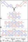
Figure 1
Figure 1
Design of RNA molecules for paranemic assembly. The cohesion of two paranemic strands designed to interact over (A) 3 half-turns (36M1 and 36M2), (B) 5 half-turns (5751 and 5752), and (C) 7 half-turns (7751 and 7752). In panels A to C, “M” indicates the major groove and “m” the minor groove. (D) Correspondence between designations used for major and minor grooves spanned by cross-overs in DNA and RNA paranemic complexes. The major groove is designated “W” in DNA and “M” in RNA paranemics, while the minor groove is designated “N” in DNA and “m” in RNA. A half-turn corresponds to between 5 and 6 basepairs. Basepairs are indicated by vertical lines connecting bases on paired RNA strands. The sugar-phosphate backbone of each strand is indicated by the thick red or blue lines.
Figure 2
Figure 2
Native gel electrophoresis assay of formation of paranemic RNA complexes with 3, 5, and 7 half-turns (3HT, 5HT and 7HT complexes). Lanes 1 and 2 are size markers 3HT and 4HT. Concentrations of unlabeled molecules are given in μM units. The radio-labeled strand in each lane is indicated by (*) and is present at ~0.5 nM. Each series of experiments is separated by vertical bars. The first lane in each series contains only the labeled monomer strand. The monomer bands are indicated with “m”, dimers with “d”, and multimers by “M”.
Figure 3
Figure 3
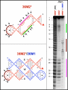
Pb2+ probing of 36M2* in the monomer form and complexed with 36M1. (Right) Denaturing gel analysis of Pb2+ probing of radiolabeled 36M2 (10 nM) in the monomer state and complexed to 36M1 (0.3 μM). “OH” indicates alkali-treated RNA. (Left) Arrows indicate Lead(II)-induced cleavages within molecule 36M2 in the monomer state (upper left) and complexed to 36M1 (lower left). The red star indicates the labeled 3’-end of 36M2.
Figure 4
Figure 4
Kinetic exchange of 36M1/36M2 complexes. (A) Schematic representation of the kinetic exchange which results from dissociation of the pre-formed, unlabeled complex composed of 36M1 (in red) and 36M2 (in light blue) and formation of the labeled complex composed of 36M1 bound to labeled 36M2* (dark blue), displacing unlabeled 36M2 (light blue). (B Left) Radiolabeled 36M2* (0.5 nM) was added (at time = 0) to the pre-formed complex of unlabeled 36M2 (0.4 μM) and 36M1 (0.3 μM). Aliquots were withdrawn at the indicated times and frozen immediately on dry ice. Samples were thawed and loaded on native gels for analysis in reverse order. (B Right) The data from two separate gels were combined and fitted with a single exponentially decaying curve, giving the exchange half-time t1/2= 100 ± 12 minutes.
Figure 5
Figure 5
Molecule 5651 assembles paranemically with 5652 but not 5552. Molecules 5651 and 5652 form six basepairs in the major groove, whereas 5552 can only form five. (Upper panel) Lanes 1 and 2: size markers; lanes 3 to 5: 5651* (0.5 nM) alone; with 5652 (1.0μM); with 5552 (1.0μM). (Lower panel) The sequences of 5552 and 5652 are compared. Red letters indicate insertions; dark blue letters indicate the terminal UUCG tetraloops.
Figure 6
Figure 6
Specificity of paranemic assembly for the 5HT system. (A) Point mutations introduced in 5752 to test binding specificity. In 5752b one substitution is present, in 5752c two substitutions are present in the same groove, and in 5752d two substitutions are present in different grooves. (B) Sequence specificity assembly experiments in which unlabeled 5751 is mixed with labeled 5752b*, 5752c* and 5752d*.
Figure 7
Figure 7
Specificity of RNA paranemic assembly with 3HT molecules. (Left) Native PAGE gel shows that cognate paranemic molecules 36M1+36M2, 36M1a+36M2a, and 36M1b+36M2b, all form dimer complexes, while non-cognate pairs 36M1a+36M2, 36M1a+36M2b, 36M1b+36M2, and 36M1b+36M2a, do not. Radiolabeled molecules are indicated by * and are at 0.5nM, while unlabeled molecules are at 50 nM. (Right) the mutations (indicated by colors) introduced in 36M1 and 36M2 to test the specificity of binding. Molecules 36M1a and 36M2a contain compensating mutations, as do 36M1b and 36M2b.
Figure 8
Figure 8
Effect of a groove size on paranemic assembly with 3HT molecules. (A) Native PAGE gel shows that only combinations of 38M1+38M2, 37M1+37M2, 35M1+35M2 and 37M1+38M2, form paranemic dimer complexes. Radiolabeled molecules are indicated by * and are at 0.5nM, while unlabeled molecules are at 200 nM. (B) Sequences of all molecules used for this experiment. Red letters indicate insertion mutations. Red gaps indicate deletion mutations.
Figure 9
Figure 9
Determination of the dissociation constants (Kd's) for 35M1/35M2, 36M1/36M2, and 37M1/37M2 dimers. (A) Schematic representation of the assembly process for 36M1 and radiolabeled 36M2* paranemic molecules. (B) Radiolabeled 35M2*, 36M2*, and 37M2* (~0.5nM) were titrated with increasing amounts on 35M1, 36M1, and 37M1 (indicated in _M units). C: Quantification of gel data for Kd determinations, each represents combined fitting of 2 datasets. The Axes are marked in units of nM.

References

    1. Ball P. Nanotechnology. 2002;13:R15–R28.
    1. Seeman NC, Belcher AM. Proc Natl Acad Sci U S A. 2002;99(Suppl 2):6451–5. - PMC - PubMed
    1. Goodsell DS. Bionanotechnology: Lessons from Nature. Wiley-Liss; Hoboken, New Jersey: 2004.
    1. Jaeger L, Leontis NB. Angew Chem Int Ed Engl. 2000;39:2521–2524. - PubMed
    1. Jaeger L, Westhof E, Leontis NB. Nucleic Acids Res. 2001;29:455–63. - PMC - PubMed

Publication types