

Impact of glucose-dependent insulinotropic peptide on age-induced bone loss
- PMID: 18072880
- PMCID: PMC2669161
- DOI: 10.1359/jbmr.071202
Impact of glucose-dependent insulinotropic peptide on age-induced bone loss
Abstract
GIP is an important hormonal link between nutrition and bone formation. We show for the first time that BMSCs express functional GIP receptors, that expression decreases with aging, and that elevations in GIP can prevent age-associated bone loss.
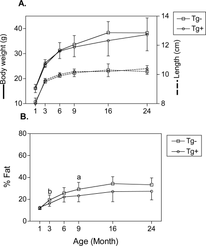
Introduction: We previously showed that C57BL/6 mice lose bone mass as they age, particularly between 18 and 24 mo of age. The mechanisms involved in this age-dependent induced bone loss are probably multifactorial, but adequate nutrition and nutritional signals seem to be important. Glucose-dependent insulinotropic peptide (GIP) is an enteric hormone whose receptors are present in osteoblasts, and GIP is known to stimulate osteoblastic activity in vitro. In vivo, GIP-overexpressing C57BL/6 transgenic (GIP Tg(+)) mice have increased bone mass compared with controls. Bone histomorphometric data suggest that GIP increases osteoblast number, possibly by preventing osteoblastic apoptosis. However, potential GIP effects on osteoblastic precursors, bone marrow stromal cells (BMSCs), had not previously been examined. In addition, effects of GIP on age-induced bone loss were not known.
Materials and methods: Changes in BMD, biomechanics, biomarkers of bone turnover, and bone histology were assessed in C57BL/6 GIP Tg(+) versus Tg(-) (littermate) mice between the ages of 1 and 24 mo of age. In addition, age-related changes in GIP receptor (GIPR) expression and GIP effects on differentiation of BMSCs were also assessed as potential causal factors in aging-induced bone loss.
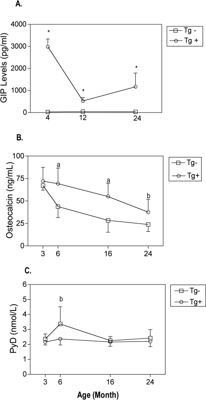
Results: We report that bone mass and bone strength in GIP Tg(+) mice did not drop in a similar age-dependent fashion as in controls. In addition, biomarker measurements showed that GIP Tg(+) mice had increased osteoblastic activity compared with wildtype control mice. Finally, we report for the first time that BMSCs express GIPR, that the expression decreases in an age-dependent manner, and that stimulation of BMSCs with GIP led to increased osteoblastic differentiation.
Conclusions: Our data show that elevated GIP levels prevent age-related loss of bone mass and bone strength and suggest that age-related decreases in GIP receptor expression in BMSCs may play a pathophysiological role in this bone loss. We conclude that elevations in GIP may be an effective countermeasure to age-induced bone loss.
Figures


References
-
- Bollag RJ, Zhong Q, Ding KH, Phillips P, Zhong L, Qin F, Cranford J, Mulloy AL, Cameron R, Isales CM. Glucose-dependent insulinotropic peptide is an integrative hormone with osteotropic effects. Mol Cell Endocrinol. 2001;177:35–41. - PubMed
-
- Bollag RJ, Zhong Q, Phillips P, Min L, Zhong L, Cameron R, Mulloy AL, Rasmussen H, Qin F, Ding KH, Isales CM. Osteoblast-derived cells express functional glucose-dependent insulinotropic peptide receptors. Endocrinology. 2000;141:1228–1235. - PubMed
-
- Zhong Q, Itokawa T, Sridhar S, Ding KH, Xie D, Kang B, Bollag WB, Bollag RJ, Hamrick M, Insogna K, Isales CM. Effects of glucose-dependent insulinotropic peptide on osteoclast function. Am J Physiol Endocrinol Metab. 2007;292:543–548. - PubMed
-
- Xie D, Zhong Q, Ding KH, Cheng H, Williams S, Correa D, Bollag WB, Bollag RJ, Insogna K, Troiano N, Coady C, Hamrick M, Isales CM. Glucose-dependent insulinotropic peptide-overexpressing transgenic mice have increased bone mass. Bone. 2007;40:1352–1360. - PubMed
-
- Tsukiyama K, Yamada Y, Yamada C, Harada N, Kawasaki Y, Ogura M, Bessho K, Li M, Amizuka N, Sato M, Udagawa N, Takahashi N, Tanaka K, Oiso Y, Seino Y. Gastric inhibitory polypeptide as an endogenous factor promoting new bone formation after food ingestion. Mol Endocrinol. 2006;20:1644–1651. - PubMed
Publication types
MeSH terms
Substances
Grants and funding
LinkOut - more resources
Full Text Sources
Other Literature Sources
Medical
Molecular Biology Databases
Miscellaneous
