. 2008 Feb;36(2):666-75.
doi: 10.1093/nar/gkm1080. Epub 2007 Dec 10.

Epstein-Barr virus-encoded microRNA miR-BART2 down-regulates the viral DNA polymerase BALF5

Affiliations

Epstein-Barr virus-encoded microRNA miR-BART2 down-regulates the viral DNA polymerase BALF5

Stephanie Barth et al. Nucleic Acids Res. 2008 Feb.

Abstract

MicroRNAs (miRNAs) have been implicated in sequence-specific cleavage, translational repression or deadenylation of specific target mRNAs resulting in post-transcriptional gene silencing. Epstein-Barr virus (EBV) encodes 23 miRNAs of unknown function. Here we show that the EBV-encoded miRNA miR-BART2 down-regulates the viral DNA polymerase BALF5. MiR-BART2 guides cleavage within the 3'-untranslated region (3'UTR) of BALF5 by virtue of its complete complementarity to its target. Induction of the lytic viral replication cycle results in a reduction of the level of miR-BART2 with a strong concomitant decrease of cleavage of the BALF5 3'UTR. Expression of miR-BART2 down-regulates the activity of a luciferase reporter gene containing the BALF5 3'UTR. Forced expression of miR-BART2 during lytic replication resulted in a 40-50% reduction of the level of BALF5 protein and a 20% reduction of the amount of virus released from EBV-infected cells. Our results are compatible with the notion that EBV-miR-BART2 inhibits transition from latent to lytic viral replication.


Figures

Figure 1.
Figure 1.
(A) Genomic localization of the EBV miRNAs. The top row of the schematic shows the location of the genes expressed in latently infected B-cells. The position of the three clusters of the miRNAs within the genome is shown in greater detail in the bottom. The deletion within the B95.8 virus isolate is indicated. For the exact location of the EBV-miRNAs, refer to references 15–17. (B) Schematic representation of luciferase reporters containing the 3′ untranslated region (Luc-BALF5-3′UTR) of the EBV BALF5 DNA polymerase gene. The 3′UTR of BALF5 extends in leftward orientation on the EBV genome from nucleotides 153228 to 152188. The potential binding site of EBV miR-BART2 (encoded on the opposite strand) extends from 152768 to 152746 and was deleted by PCR in the reporter Luc-BALF5 3′UTR-mut. Numbering refers to gene bank accession number AJ507799. Note that the drawing is not to scale.
Figure 2.
Figure 2.
miR-BART2 down-regulates the BALF5 3′UTR. (A) Northern blot detection of ectopically expressed miR-BART2 and miR-155 using the indicated probes. RNA extracted from HeLa cells 48 h after transfection with the vector pSG5-miR-BART2 (lane designated ‘HeLa+miR-BART2’) was analysed in parallel with RNA from BL41 and B95.8 cells (EBV-negative and -positive, respectively). Total RNA of HeLa cells either transfected or untransfected with pSG5-mir-155 was analysed by northern blotting for the expression of miR-155. The positions of the precursor and the mature miRNA are indicated. (B) Effect of miR-BART2 on the BALF5 3′UTR. miR-BART2 and the Luc-BALF5-3′UTR reporter were co-expressed in the indicated combinations. The activity obtained with the reporter alone was set to 100%. Graph B represents the mean values of six independent experiments carried out in duplicate (±SEM). (C) miR-BART2 and a luciferase reporter containing the BALF5 3′UTR with a deletion of the BART2-recognition site were co-expressed in the indicated combinations and analysed as in (B). (D) Effect of miR-BART2 on the parental (empty) vector pGL3-promoter (‘Luc’). MiR-BART2 and the pGL3-reporter were co-expressed in the indicated combinations. The activity obtained with the reporter alone was set to 100%. The statistical analysis showed an insignificant effect (P = 0.257). (E) miR-BART2 and a luciferase reporter containing the LMP2A 3′UTR were co-expressed in the indicated combinations and analysed as in (B). The activity obtained with the reporter alone was set to 100%. (F) Effect of miR-155 on the pGL3-BALF5 3′-UTR reporter; the reporter alone was set to 100%. The statistical analysis showed an insignificant effect (P = 0.092). Graphs C, D, E and F represent the mean values of four independent experiments carried out in duplicate (±SEM).
Figure 3.
Figure 3.
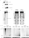
miR-BART2-directed cleavage of BALF5 mRNA is reduced during lytic replication. (A) Immunoprecipitation analysis of Ago2 using monoclonal antibody 3C7. Extracts of 293-T cells expressing HA-tagged Ago2 were subjected to precipitation using the Ago2-specific rat monoclonal antibody 3C7 (IgG1) or isotype control antibody. Whole-cell extract containing HA-Ago2 was co-electrophoresed in the adjacent lane designated ‘input’. After transfer to nitrocellulose membrane, the precipitated Ago2 was visualized using 3C7. The position of rat immunoglobulin heavy and light chains are indicated. Molecular mass marker proteins (×10−3 kDa) were, in descending order: phosphorylase B, bovine serum albumin, ovalbumin, carboanhydrase. (B) Ago2-mediated cleavage of RNA. Cytoplasmic extracts generated from the indicated cell lines with or without previous TPA treatment were subjected to immunoprecipitation using anti-Ago2 antibodies. Immunoprecipitates were subsequently incubated with 32P-cap-labelled RNAs, which contained perfect complementary sequences to the viral miRNA BART2. The cleaved RNA products were separated by 8% denaturing RNA–PAGE. Lanes denoted ‘T1’ show nuclease T1 digestions of the RNA substrates. (C) Ago-2 mediated cleavage of RNA. Extracts of the indicated cell lines with or without TPA-treatment were compared. The RNA sequences complementary to miR-BART2 or miR-19b are indicated by black bars to both sides. Arrows denote the actual cleavage sites. (D) Detection of miRNA levels during EBV lytic replication. Total RNA from EBV-negative BL41 cells, from type-2 EBV-infected Jijoye cells and from type-1 EBV infected B95.8 cells either treated (+TPA) or not treated (−TPA) with tetradecanoylphorbol acetate (TPA), was assayed in a northern blot with the indicated 32P-labelled probes. The loading control (tRNA) is shown below each blot.
Figure 4.
Figure 4.
Reduced repression (‘De-repression’) of the BALF5-3′UTR during lytic cycle replication. The pGL3 vector containing the BALF5-3′UTR (A) or the reporter with the mutated BALF5-3′UTR (B) were transfected alone or with the p509 vector encoding the lytic activator BZLF1 into 293-EBV cells. The luciferase activities observed without p509 were set to 100%. Activation of Luc-BALF5-3′UTR with p509 was 7.7-fold (P = 0.003); activation for pGL3 with the mutated binding site was 1.6-fold (P = 0.07). Graphs represent the mean value of four (A) or rather eight (B) independent assays carried out in duplicate (±SEM).
Figure 5.
Figure 5.
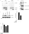
miR-BART2 down-regulates BALF5 polymerase. (A) Identification of BALF5 protein using the monoclonal antibody 4C12. Whole-cell extracts of B95.8 B-cells either treated (+) or untreated (−) with TPA were analysed by western blotting as shown in the left panel. The blots were stained with the novel BALF5-specific antibody 4C12, an antibody directed against β-actin as a loading control and BZLF1-specific monoclonal antibody BZ-1 to verify the induction of EBV lytic replication. Detection of BALF5 in EBV-infected 293 cells without (–) and after (+) induction of lytic replication by BZLF1 using the vector p509 (18) is shown in the right panel. (B) Immunoprecipitation of BALF5. Extract of TPA-treated Raji cells was incubated either with BALF5-specific antibody 4C12 or irrelevant isotype control as indicated. Immune complexes were collected using protein G Sepharose (Amersham-Pharmacia). The precipitated BALF5 protein was analysed in a western blot using 4C12 as primary antibody; bound antibody was visualized by the ECL method; the lanes designated ‘Raji’, shows whole-cell extract prior to precipitation. (C) Reduction of BALF5 protein levels by miR-BART2. 293-EBV cells were transfected with BZLF1 expression vector p509 in combination with miR-BART2 expression vector or pSG5 control. BALF5 protein was stained using the monoclonal antibody 4C12, β-actin served as a loading control (left panel); statistical analysis of the BALF5 protein reduction by miR-BART2. The amount of BALF5 protein with or without BART2 expression from three independent assays as shown in (C) was determined and statistically analysed. The reduction of 30–40% after co-expression of miR-BART2 was statistically significant (right panel; P = 0.0037). (D) Reduction in virus load by miR-BART2. Viral replication in 293-EBV cells was induced by expression of BZLF1 using the vector p509. The amount of virus released was determined by quantitative real-time PCR. The value obtained by co-transfection of the empty control vector pSG5 was set to 100%. Co-expression of miR-BART2 resulted in a statistically significant reduction of the virus load by 20% (P = 0.039), co-expression of miR-155 resulted in a non-significant reduction by 5–10% (P = 0.155).

References

    1. Bartel DP. MicroRNAs: genomics, biogenesis, mechanism, and function. Cell. 2004;116:281–297. - PubMed
    1. Ambros V. The functions of animal microRNAs. Nature. 2004;431:350–355. - PubMed
    1. Pillai RS, Bhattacharyya SN, Filipowicz W. Repression of protein synthesis by miRNAs: how many mechanisms? Trends Cell Biol. 2007;17:118–126. Epub 2007 Jan 2002. - PubMed
    1. Borchert GM, Lanier W, Davidson BL. RNA polymerase III transcribes human microRNAs. Nat. Struct. Mol. Biol. 2006;12:12. - PubMed
    1. Cai X, Hagedorn CH, Cullen BR. Human microRNAs are processed from capped, polyadenylated transcripts that can also function as mRNAs. RNA. 2004;10:1957–1966. Epub 2004 Nov 1953. - PMC - PubMed

Publication types

MeSH terms