. 2008 Mar;93(3):1020-9.
doi: 10.1210/jc.2007-2331. Epub 2007 Dec 11.

Protein kinase A-independent inhibition of proliferation and induction of apoptosis in human thyroid cancer cells by 8-Cl-adenosine

Affiliations

Protein kinase A-independent inhibition of proliferation and induction of apoptosis in human thyroid cancer cells by 8-Cl-adenosine

Audrey J Robinson-White et al. J Clin Endocrinol Metab. 2008 Mar.

Abstract

Purpose: Protein kinase A (PKA) affects cell proliferation in many cell types and is a potential target for cancer treatment. PKA activity is stimulated by cAMP and cAMP analogs. One such substance, 8-Cl-cAMP, and its metabolite 8-Cl-adenosine (8-Cl-ADO) are known inhibitors of cancer cell proliferation; however, their mechanism of action is controversial. We have investigated the antiproliferative effects of 8-Cl-cAMP and 8-CL-ADO on human thyroid cancer cells and determined PKA's involvement.

Experimental design: We employed proliferation and apoptosis assays and PKA activity and cell cycle analysis to understand the effect of 8-Cl-ADO and 8-Cl-cAMP on human thyroid cancer and HeLa cell lines.

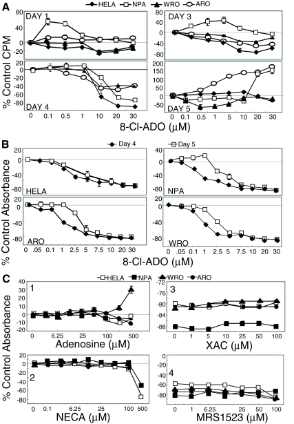
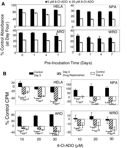
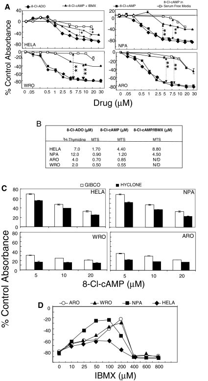
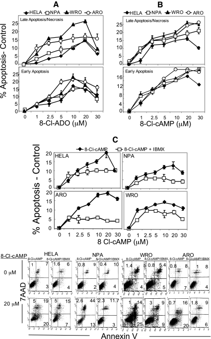
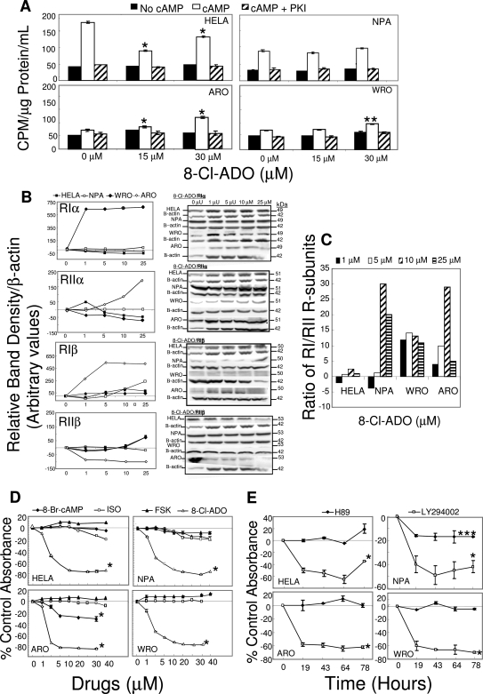
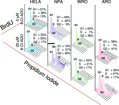
Results: 8-Cl-ADO inhibited proliferation of all cells, an effect that lasted for at least 4 d. Proliferation was also inhibited by 8-Cl-cAMP, but this inhibition was reduced by 3-isobutyl-1-methylxanthine; both drugs stimulated apoptosis, and 3-isobutyl-1-methylxanthine drastically reduced 8-Cl-cAMP-induced cell death. 8-Cl-ADO induced cell accumulation in G1/S or G2/M cell cycle phases and differentially altered PKA activity and subunit levels. PKA stimulation or inhibition and adenosine receptor agonists or antagonists did not significantly affect proliferation.

Conclusions: 8-Cl-ADO and 8-Cl-cAMP inhibit proliferation, induce cell cycle phase accumulation, and stimulate apoptosis in thyroid cancer cells. The effect of 8-Cl-cAMP is likely due to its metabolite 8-Cl-ADO, and PKA does not appear to have direct involvement in the inhibition of proliferation by 8-Cl-ADO. 8-Cl-ADO may be a useful therapeutic agent to be explored in aggressive thyroid cancer.


Figures

Figure 1
Figure 1
8-Cl-ADO inhibits [3H]thymidine uptake and cell metabolism in thyroid cancer cells and in HeLa cells, with no effect on adenosine receptors. A, 8-Cl-ADO inhibits [3H]thymidine uptake in a time- and concentration-dependent manner. Cells in 24-well culture plates were incubated for 1–5 d with increasing 8-Cl-ADO concentrations (0–30 μm), in the presence of 2 μCi [3H]thymidine per well. Counts per minute (CPM) were determined by scintillation count, and values were calculated as percent of control counts per minute. One representative experiment is shown. B, Cells in 96-well culture plates were incubated for 4 and 5 d with 0–30 μm 8-Cl-ADO. C, Cells in 96-well culture plates were incubated for 4 d with adenosine (0–500 μm, panel 1), NECA (0–500 μm, panel 2), XAC (0–100 μm) plus 20 μm 8-Cl-ADO or 8-Cl-ADO alone (panel 3), and MRS1523 (0–100 μm) plus 20 μm 8-Cl-ADO, or 8-Cl-ADO alone (panel 4). In B and C, Cell Titer 96 AQ solution was added 3.5 h before plates were read using an ELISA reader at 460 nm. Results in B and C are percentage of control absorbance. In C (panels 3 and 4), zero represents 20 μm 8-Cl-ADO alone. Points are mean ± sem of four wells (A), mean ± sem of four experiments (B), and mean ± sem of three experiments (C); P < 0.0001 (B).
Figure 2
Figure 2
Loss of inhibition with 8-Cl-ADO with prolonged incubation time: intracellular drug metabolism. A, 8-Cl-ADO (5 and 20 μm) was preincubated (minus cells) for 0–7 d at 37 C before its addition to cells for an additional 4 d; B, cells in 24-well culture plates were incubated with 8-Cl-ADO (20 μm) and [3H]thymidine (2 μCi per well) for 4 and 5 d. In some cultures, 8-Cl-ADO was replenished on d 4 and cultures assayed on d 5. Levels of intranuclear counts per minute (CPM) were determined by scintillation count, and values expressed as percent of control counts per minute. In A, Cell Titer 96 AQ solution was added 3.5 h before plates were read using an ELISA plate reader. Results are expressed as percentage of control absorbance. Points are mean ± sd of two experiments (A) and mean ± sem of three experiments (B); *, P = 0.032 (A); **, P < 0.0001 (B).
Figure 3
Figure 3
Metabolism of 8-Cl-cAMP to 8-Cl-ADO: effect of IBMX and serum-free media on extracellular drug metabolism. A, Cells in 96-well culture plates were incubated with increasing concentrations (0–30 μm) of 8-Cl-ADO, 8-Cl-cAMP, or 8-Cl-cAMP plus IBMX (50 μm) in media containing 10% FBS (Hyclone). Cells were also incubated with 8-Cl-cAMP in serum-free medium. All cultures were incubated for 4 d. B, List of IC50 values from [3H]thymidine and MTS assays. C, Cells in 96-well culture plates were incubated (4 d) with increasing concentrations of 8-Cl-cAMP in media containing serum from GIBCO/Invitrogen or from Hyclone. D, Cells in 96-well culture plates were incubated (4 d) with increasing concentrations of IBMX (0–800 μm) and/or 8-Cl-cAMP (25 μm). In A, C, and D, Cell Titer 96 AQ solution was added for 3.5 h before plates were read using an ELISA plate reader at 460 nm. Results are expressed as percentage of control absorbance and are mean ± sem of six experiments (A) and mean ± sd of two experiments (C and D). *, P = 0.01; **, P < 0.0001 (A).
Figure 4
Figure 4
8-Cl-cAMP and 8-Cl-ADO stimulate apoptosis in HeLa cells and in thyroid cancer cells. IBMX inhibits 8-Cl-cAMP-induced apoptosis. Cells were incubated with increasing concentrations of 8-Cl-ADO (A), 8-Cl-cAMP (B), or 8-Cl-cAMP plus (50 μm) IBMX (C). Apoptosis was assessed by annexin V-FITC/7AAD assay staining and measured by FACScalibur flow cytometry. Points are mean ± sem of three experiments (A), four experiments (B), and three experiments (C), and results are expressed as percentage of apoptosis (minus apoptosis in untreated cells). Dot plots in C are of one representative experiment. P = 0.004 to <0.0001 (C).
Figure 5
Figure 5
Differential effect of 8-Cl-ADO on PKA activity in thyroid cancer cells and in HeLa cells: no significant effect of PKA pathway stimulants or the PKA activity inhibitor H89 on cell metabolism/proliferation. A, Cells in 175-cm2 flasks were incubated with 8-Cl-ADO (15 and 30 μm) for 4 d, and cell extracts were exposed to cAMP or to cAMP plus the PKA-specific inhibitor PKI. PKA activity levels were then determined. Results are expressed as cpm [γ32P]dATP per microgram protein per milliliter and are mean ± sem of three experiments. B, Cells in 175-cm2 flasks were incubated with 8-Cl-ADO (0–25 μm) for 4 d and lysed, and gel electrophoresis and immunoblot assay were performed using antibodies to PKA R-subunits. C, Ratio of PKA R-subunits RIα plus RIβ/ RIIα plus RIIβ from B above. D, Cells in 96-well culture plates were incubated for 4 d with increasing concentrations (0–40 μm) of the PKA pathway stimulants 8-Br-cAMP, isoproterenol (ISO), and forskolin (FSK), or with 8-Cl-ADO. E, cells were incubated with the PKA pathway inhibitor, H89 (50 nm) or with the PKB (Akt)-pathway inhibitor LY294002 (30 μm) for 0–78 h. Cell titer 96 AQ solution was added to cultures in D and E for 3.5 h, before plates were read using an ELISA plate reader at 460 nm. Points in B are mean ± sem of three experiments, expressed as percentage of control band density/Β-actin (arbitrary values). Bands are representative of one experiment. Results are expressed as percentage of control absorbance ± sem of three experiments (D) or mean ± sd of two experiments (E). *, P < 0.0001 (A, D, and E); **, P = 0.0005 (A); ***, P = 0.01 (E) compared with baseline.
Figure 6
Figure 6
8-Cl-ADO induces the accumulation of cells in G1/S and G2/M phases of the cell cycle. Cells, synchronized (72 h) in low-serum (0.1%) media were incubated for 3 d with or without 8-Cl-ADO (20 μm) in complete medium. Cells were pulse labeled with BrdU, followed by the addition of anti-BrdU-FITC mAb and propidium iodide. Cell cycle analysis was determined by FACScalibur flow cytometry, using Cell Quest software. On the y-axis is the percentage of newly formed DNA (BrdU) content as indicated by anti-BrdU-FITC mAb in each cell cycle phase on the x-axis is propidium iodide. Three-dimensional plots are from one representative experiment.

References

    1. Sakamoto A 2004 Definition of poorly differentiated carcinoma of the thyroid: the Japanese experience. Endocr Pathol 15:307–311 - PubMed
    1. Nikiforov YE 2004 Genetic alterations involved in the transition from well-differentiated to poorly differentiated and anaplastic thyroid carcinomas. Endocr Pathol 15:319–327 - PubMed
    1. Delellis RA, Lloyd RV, Hertz PU, Eng C 2004 Tumors of the thyroid and parathyroid. In: Delellis RA, Lloyd RV, Hertz PU, Eng C, eds. WHO classification of tumors: pathology and genetics. Lyon, France: IARC Press; 51–80
    1. Liska J, Altanerova V, Galbavy S, Stvrtina S, Brtko J 2005 Thyroid tumors: histological classification and genetic factors involved in the development of thyroid cancer. Endocr Res 39:73–83 - PubMed
    1. Ain KB 1998 Anaplastic thyroid carcinoma: behavior, biology and therapeutic approaches. Thyroid 8:715–726 - PubMed

Publication types

MeSH terms

Substances