. 2007 Dec;177(4):2293-307.
doi: 10.1534/genetics.107.074963.

The reacquisition of biotin prototrophy in Saccharomyces cerevisiae involved horizontal gene transfer, gene duplication and gene clustering

Affiliations

The reacquisition of biotin prototrophy in Saccharomyces cerevisiae involved horizontal gene transfer, gene duplication and gene clustering

Charles Hall et al. Genetics. 2007 Dec.

Abstract

The synthesis of biotin, a vitamin required for many carboxylation reactions, is a variable trait in Saccharomyces cerevisiae. Many S. cerevisiae strains, including common laboratory strains, contain only a partial biotin synthesis pathway. We here report the identification of the first step necessary for the biotin synthesis pathway in S. cerevisiae. The biotin auxotroph strain S288c was able to grow on media lacking biotin when BIO1 and the known biotin synthesis gene BIO6 were introduced together on a plasmid vector. BIO1 is a paralog of YJR154W, a gene of unknown function and adjacent to BIO6. The nature of BIO1 illuminates the remarkable evolutionary history of the biotin biosynthesis pathway in S. cerevisiae. This pathway appears to have been lost in an ancestor of S. cerevisiae and subsequently rebuilt by a combination of horizontal gene transfer and gene duplication followed by neofunctionalization. Unusually, for S. cerevisiae, most of the genes required for biotin synthesis in S. cerevisiae are grouped in two subtelomeric gene clusters. The BIO1-BIO6 functional cluster is an example of a cluster of genes of "dispensable function," one of the few categories of genes in S. cerevisiae that are positionally clustered.


Figures

F<sc>igure</sc> 1.—
Figure 1.—
Diagram of the biotin biosynthesis pathway in S. cerevisiae. (A) Genomic position of the six known genes involved in biotin biosynthesis and intermediate transport. The chromosomal positions of BIO1/BIO6 genes are based on strain S288C and represent duplicated regions on chromosomes I and VIII. (B) Position of the genes in the biotin biosynthesis pathway. The biochemical intermediates in this pathway are based on work in E. coli, Bacillus sp., and S. cerevisiae. The precursor of pimeloyl-CoA is malonyl-CoA in E. coli and pimelic acid in Bacillus sp.
F<sc>igure</sc> 2.—
Figure 2.—
Phylogeny of BIO1 from S. cerevisiae and related genes and pimeloyl-CoA synthetase genes from prokaryotes (left) and species phylogeny based on SSU rDNA (right). BIO1 (clade a) is a duplicate copy of YJR154W. The pimeloyl-CoA synthetase activity of the genes in species in boldface type has been experimentally verified (Streit and Entcheva 2003). Pimeloyl-CoA synthetase activity appears to be highly divergent among the prokaryotes with some bacteria requiring the two-gene bioCbioH complex (clades b and e) and others requiring a single gene such as bioW or bioZ (clades c and d). Trees were constructed by NJ in ClustalX. Numbers indicate bootstrap support for each node of 1000 replicates. Taxa marked with asterisks indicate putative phytanoyl-CoA dioxygenases.
F<sc>igure</sc> 3.—
Figure 3.—
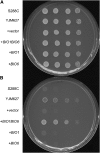
Growth of S. cerevisiae strains on media containing 2 μg/liter biotin (A) and lacking biotin (B). Cells were grown overnight in YPD, transferred to liquid minimal media lacking biotin, grown overnight, transferred a second time to liquid minimal media lacking biotin, and then spotted onto plates. S288c is a biotin auxotroph. YJM627 is a biotin prototroph. +vector indicates vector alone (plasmid PPH001), +BIO1 includes BIO1 gene (plasmid PCH001), +BIO6 includes BIO6 gene (plasmid PCH002), and +BIO1BIO6 includes both genes on a single plasmid (plasmid PCH003).
F<sc>igure</sc> 4.—
Figure 4.—
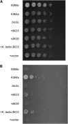
Growth of S. cerevisiae strains on media containing 2 μg/liter biotin (A) and lacking biotin (B). Cells were grown overnight in YPD, transferred to liquid minimal media lacking biotin, grown overnight, transferred a second time to liquid minimal media lacking biotin, and then spotted onto plates. S288c is a biotin auxotroph. A364a is a biotin prototroph. All plasmids were transformed into strain SCCH015 (A364a derived, bio1∷HygB). +vector indicates vector alone (plasmid p427-TEF), +BIO1 includes BIO1 gene (plasmid PCH004), and +YJR154W includes the YJR154W gene (plasmid PCH005).
F<sc>igure</sc> 5.—
Figure 5.—
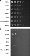
Growth of S. cerevisiae strains on media containing 2 μg/liter biotin (A) and lacking biotin (B). Cells were grown overnight in YPD, transferred to liquid minimal media lacking biotin, grown overnight, transferred a second time to liquid minimal media lacking biotin, and then spotted onto plates. S288c is a biotin auxotroph. A364a is a biotin prototroph. All plasmids were transformed into strain SCCH016 (A364a derived, bio3∷HygB). +vector indicates vector alone (p427-TEF), +BIO3 and +BIO3 K. lactis includes the BIO3 gene from S. cerevisiae and K. lactis, respectively (plasmids PCH006 and PCH007), and +BIO6 includes the BIO6 gene (plasmid PCH008).
F<sc>igure</sc> 6.—
Figure 6.—
Growth of S. cerevisiae strains on media containing 2 μg/liter biotin (A) and lacking biotin (B). Cells were grown overnight in YPD, transferred to liquid minimal media lacking biotin, grown overnight, transferred a second time to liquid minimal media lacking biotin, and then spotted onto plates. S288c is a biotin auxotroph. A364a is a biotin prototroph. All plasmids were transformed into strain SCCH017 (A364a derived, bio6∷HygB). +vector indicates vector alone (p427-TEF), +BIO3 and +BIO3 K. lactis includes the BIO3 gene from S. cerevisiae and K. lactis, respectively (plasmids PCH006 and PCH007), and +BIO6 includes the BIO6 gene (plasmid PCH008).
F<sc>igure</sc> 7.—
Figure 7.—
Variability of biotin biosynthesis in hemiascomycete fungi. Strains of Candida albicans (2), K. lactis (2), S. cerevisiae (37), S. paradoxus (35), and S. kluyveri (2) were replica plated from minimal media onto media lacking biotin. Some strains show no growth, some weak growth, and others robust growth. This experiment was repeated three times. Growth indicated is an average for all three experiments. It is likely that growth differences are due to copy number differences in BIO6 and BIO1 (Wu et al. 2005). Strains used and their relative growth on minimal media lacking biotin are summarized in Table 1.
F<sc>igure</sc> 8.—
Figure 8.—
Phylogeny of KAPA synthetase (bioF and BIO6) and DAPA synthetase (bioA and BIO3) indicates horizontal gene transfer of BIO3 into fungi from proteo bacteria (A) and gain of KAPA synthetase function by duplication of BIO3 to form BIO6 (B). Gene phylogeny KAPA synthetase is shown in a and DAPA synthetase in b. Species phylogeny was determined by ribosomal small subunit (SSU). Both trees were determined by distance methods. The numbers indicate bootstrap support for each node of 1000 replicates.
F<sc>igure</sc> 9.—
Figure 9.—
Phylogeny of dethiobiotin synthetase (bioD and BIO4) indicates horizontal gene transfer of BIO4 into fungi from bacteria. Gene phylogeny of dethiobiotin synthetase (left) and species phylogeny (right) were determined by ribosomal small subunit (SSU). Both trees were determined by distance methods. The numbers indicate bootstrap support for each node of 1000 replicates.
F<sc>igure</sc> 10.—
Figure 10.—
Loss of the eukaryotic biotin biosynthesis pathway in the hemiascomycete lineage and reconstruction by horizontal gene transfer and gene duplication. Phylogram represents the evolutionary relationship between fungi. Rows indicate enzyme type. E indicates eukaryotic-type enzyme and P indicates prokaryotic-type enzyme. D indicates genes formed by duplication and neofunctionalization. Question marks indicate that the gene responsible for this step is unknown. Columns indicate enzyme function. PCAS indicates pimeloyl-CoA synthetase, KAPAS indicates KAPA synthetase, DAPAS indicates DAPA synthetase, DTBS indicates dethiobiotin synthetase, and BS indicates biotin synthetase. While S. kluyveri is a biotin prototroph (Figure 8), the genome of this organism does not appear to encode either a eukaryotic or a prokaryotic type PCAS or KAPAS.

References

    1. Abe, Y., T. Suzuki, C. Ono, K. Iwamoto, M. Hosobuchi et al., 2002. Molecular cloning and characterization of an ML-236B (compactin) biosynthetic gene cluster in Penicillium citrinum. Mol. Genet Genomics 267: 636–646. - PubMed
    1. Ashburner, M., C. Ball, J. A. Blake, D. Botstein, H. Butler et al., 2000. Gene ontology: tool for the unification of biology. Nat. Genet. 25: 25–29. - PMC - PubMed
    1. Benson, D. A., M. S. Boguski, D. J. Lipman, J. Ostell, B. F. Ouellette et al., 1999. GenBank. Nucleic Acids Res. 27: 12–17. - PMC - PubMed
    1. Bower, S., J. Perkins, R. R. Yocum, P. Serror, A. Sorokin et al., 1995. Cloning and characterization of the Bacillus subtilis birA gene encoding a repressor of the biotin operon. J. Bacteriol. 177: 2572–2575. - PMC - PubMed
    1. Brachmann, C. B., A. Davies, G. J. Cost, E. Caputo, J. Li et al., 1998. Designer deletion strains derived from Saccharomyces cerevisiae S288c: a useful set of strains and plasmids for PCR-mediated gene disruption and other applications. Yeast 14: 115–132. - PubMed

MeSH terms

Associated data