. 2007 Dec 12;2(12):e1297.
doi: 10.1371/journal.pone.0001297.

Human RECQ1 is a DNA damage responsive protein required for genotoxic stress resistance and suppression of sister chromatid exchanges

Affiliations

Human RECQ1 is a DNA damage responsive protein required for genotoxic stress resistance and suppression of sister chromatid exchanges

Sudha Sharma et al. PLoS One. .

Abstract

Background: DNA helicases are ubiquitous enzymes that unwind DNA in an ATP-dependent and directionally specific manner. Unwinding of double-stranded DNA is essential for the processes of DNA repair, recombination, transcription, and DNA replication. Five human DNA helicases sharing sequence similarity with the E. coli RecQ helicase have been identified. Three of the human RecQ helicases are implicated in hereditary diseases (Bloom syndrome, Werner syndrome, and Rothmund-Thomson syndrome) which display clinical symptoms of premature aging and cancer. RECQ1 helicase is the most highly expressed of the human RecQ helicases; however, a genetic disease has yet not been linked to mutations in the RECQ1 gene, and the biological functions of human RECQ1 in cellular DNA metabolism are not known.

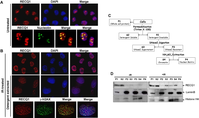
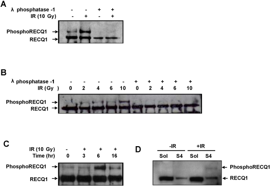
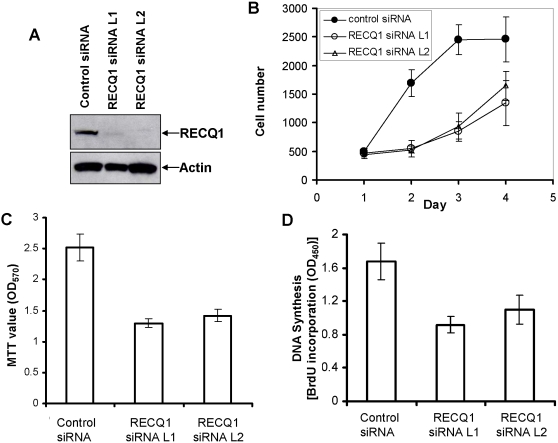
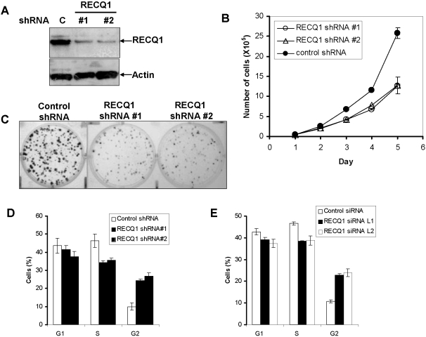
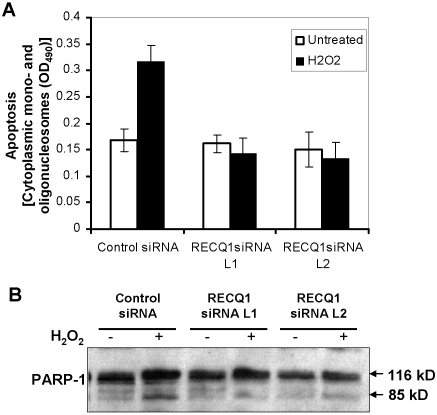
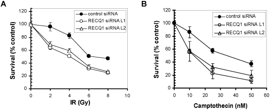
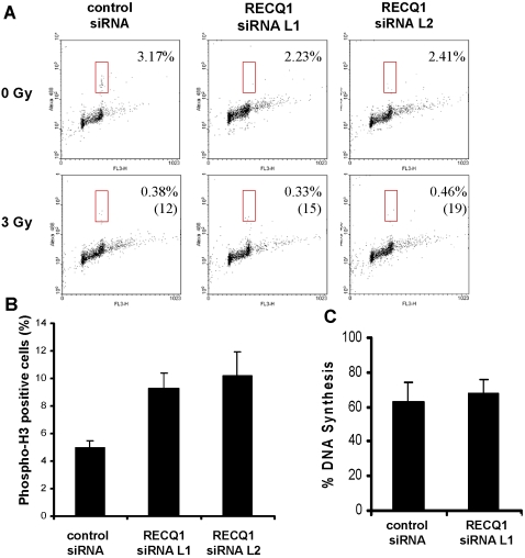
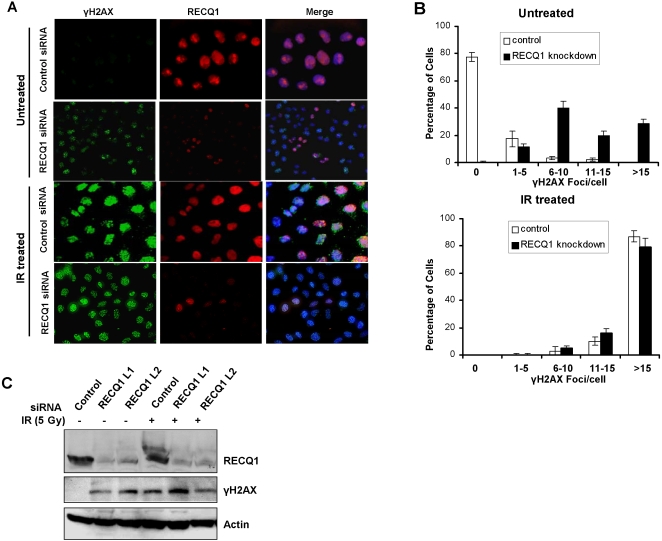
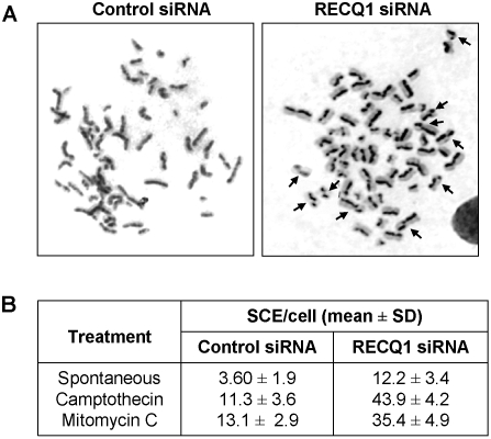
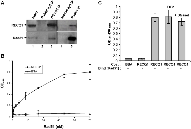
Methodology/principal findings: In this study, we report that RECQ1 becomes phosphorylated upon DNA damage and forms irradiation-induced nuclear foci that associate with chromatin in human cells. Depletion of RECQ1 renders human cells sensitive to DNA damage induced by ionizing radiation or the topoisomerase inhibitor camptothecin, and results in spontaneous gamma-H2AX foci and elevated sister chromatid exchanges, indicating aberrant repair of DNA breaks. Consistent with a role in homologous recombinational repair, endogenous RECQ1 is associated with the strand exchange protein Rad51 and the two proteins directly interact with high affinity.

Conclusion/significance: Collectively, these results provide the first evidence for a role of human RECQ1 in the response to DNA damage and chromosomal stability maintenance and point to the vital importance of RECQ1 in genome homeostasis.


Conflict of interest statement

Competing Interests: The authors have declared that no competing interests exist.

Figures

Figure 1
Figure 1. Nucleolar RECQ1 undergoes re-localization to chromatin in response to IR.
Indirect immunofluorescence was performed on HeLa cells that were either untreated or allowed to recover for 6 h from 10 Gy IR exposure as described in “Materials and Methods”. Panel A, Nucleolar localization of RECQ1 in untreated cells. Cells were co-immunostained with anti-RECQ1 and anti-nucleolin. The merged images show cells stained with RECQ1 (red) and nucleolin (green), with or without DAPI (blue). Panel B, Relocalization of RECQ1 to chromatin-bound foci following IR damage. After IR exposure, cells were stained for total RECQ1 (top panel) or in situ detergent extraction-resistant RECQ1 (middle and bottom panel). Merged images show RECQ1 (red) and DAPI (blue). Co-immunostaining of chromatin-bound RECQ1 and γ-H2AX (Panel B, bottom). Merged images show RECQ1 (red) and γ-H2AX (green), with or without DAPI (blue). Panel C, Schematic presentation of the protocol for sequential nuclear fractionation of lysates from U2OS cells either untreated or following 6 h recovery from 10 Gy IR exposure. Cytoplasmic and nucleoplasmic proteins were extracted by permeabilization with detergent, and the resulting nuclei were nuclease-digested and extracted with NH2SO4. Proteins of the supernatant (S) and pellet (P) fractions were resolved on 10% SDS-PAGE, and subsequently analyzed by Western blot for RECQ1, histone H4 (chromatin marker), and lamin B (nuclear matrix marker). Panel D, Following IR treatment, RECQ1 is enriched in the chromatin fraction (S4).
Figure 2
Figure 2. Phosphorylation of endogenous RECQ1 in response to IR.
Panel A, RECQ1 was immunoprecipitated from whole cell extracts of U2OS cells that were either untreated or allowed to recover for 6 h from 10 Gy IR exposure. The RECQ1 immunoprecipitate from each cell extract was divided in half and was either incubated or not with λ-phosphatase (500 U) for 1 h at 30°C before elution with SDS sample buffer. The immunoprecipitated proteins were resolved on 12% SDS-PAGE, transferred to PVDF membranes, and probed for RECQ1. Panel B, IR-induced RECQ1 phosphorylation is dependent on radiation dose. RECQ1 was immunoprecipitated from either untreated cells or 6 h after treatment of cells with the indicated dose of γ-radiation. Immunoprecipitated proteins were divided in two aliquots which were either treated with λ-phosphatase or left untreated. RECQ1 was detected as described above. Panel C, Time course of RECQ1 phosphorylation in response to IR. U2OS cells were subjected to γ-radiation (10 Gy), and RECQ1 was immunoprecipiated from whole cell extracts prepared at the indicated time points following IR exposure. Panel D, Phosphorylated RECQ1 is preferentially associated with chromatin in IR-treated cells. RECQ1 was immunoprecipitated from the detergent-soluble (S2) and insoluble (S4) fractions of untreated or IR treated cells, resolved on 12% SDS-PAGE, and detected by Western blot analysis.
Figure 3
Figure 3. RECQ1-depleted cells show reduced cellular proliferation.
Panel A, Small interfering RNA inhibition of RECQ1. Whole cell extracts from HeLa cells that had been transfected with control or RECQ1 siRNA (oligonucleotide L1 or L2) were subjected to Western blotting with RECQ1 or Actin antibodies (as a loading control). Panel B, Proliferation of control or RECQ1 siRNA treated cells as determined by CyQuant assay at indicated time points after transfection. Panel C, MTT assay of in vitro proliferation of control or RECQ1-siRNA transfected cells. Panel D, Colorimetric BrdU cell proliferation ELISA of control or RECQ1 siRNA treated HeLa cells. Results are taken from three independent experiments and proliferation is represented as the mean±standard deviation (SD).
Figure 4
Figure 4. Reduced cell growth and aberrant cell cycle progression of RECQ1-depleted cells.
Panel A, Short hairpin RNA (shRNA)-mediated RECQ1 depletion in HeLa cells. Western blot showing RECQ1 expression in puromycin resistant HeLa cells transfected with either control or RECQ1 shRNA (#1 or #2). Actin is used as loading control. Proliferation of control or cells transfected with RECQ1-specific shRNA plasmids was determined by Coulter counting the total number of cells at indicated time points (Panel B) and by colony forming assay (Panel C). Panels D and E, RECQ1 depletion induces G2/M accumulation. Flow cytometry was used to determine cell cycle distribution of control or RECQ1 shRNA transfected cells (Panel D) or RECQ1 siRNA transfected cells (Panel E).
Figure 5
Figure 5. Effect of RECQ1 depletion on spontaneous or oxidative stress induced apoptosis.
HeLa cells were siRNA treated for 48 h, and then incubated in complete medium for 24 h. Cells were either untreated or incubated with 400 µM H2O2 for 3 h in serum free medium, washed, and allowed to recover in complete medium for 21 h. Panel A, Enrichment of the cytoplasmic histone-associated-DNA fragments that are indicative of an ongoing apoptosis in cells with the indicated siRNA oligonucleotides. Samples were analyzed in duplicates, and data points represent the mean of three independent experiments; bars denote SD. Panel B, immunoblotting analysis of the PARP cleavage in control or RECQ1 siRNA treated HeLa cells either untreated or exposed to 400 µM H2O2.
Figure 6
Figure 6. RECQ1 depletion sensitizes cells to DNA damage induced by ionizing radiation or camptothecin.
siRNA knockdown of RECQ1 in HeLa cells leads to increased sensitivity to treatment with IR (Panel A) or CPT (Panel B). HeLa cells treated with either RECQ1 siRNA or control siRNA (#C) were plated in quadruplicate and treated with increasing doses of IR or concentrations of CPT. Total DNA content was measured as an indication of cell growth. Two independent siRNAs (#L1 and #L2) used to downregulate RECQ1 expression resulted in similar growth phenotype in HeLa cells. Three independent determinations of cell survival were performed and the mean±SD is presented.
Figure 7
Figure 7. Effect of RECQ1 depletion on G2/M and intra-S-phase DNA damage checkpoints.
Panel A, DNA damage-induced mitotic entry delay is minimally affected by the RECQ1 deficiency. HeLa cells were transfected with the indicated siRNAs and either mock-treated or exposed to 3 Gy of IR 1 h before harvesting. Mitotic cells were detected by PI and phosphohistone H3 staining and analyzed by flow cytometry. Percentages of mitotic cells and their levels normalized to control (in parentheses) are shown. Panel B, RECQ1 is involved in maintenance of IR-induced G2/M checkpoint. Cells with either control or RECQ1 siRNA were exposed to 10 Gy of IR. Nocodazole (1 µg/ml) was added to the medium at the time of IR treatment to capture cells entering mitosis. 16 h later, cells were collected for PI and phospho-histone H3 staining and analyzed by flow cytometry. Panel C, HeLa cells transfected with control or RECQ1 siRNAs were exposed to 10 Gy of IR and assayed for DNA synthesis 30 min later by [3H]thymidine incorporation. The amount of DNA synthesis after irradiation is expressed as a percentage of the level in untreated cells. Error bars indicate SD from three independent experiments.
Figure 8
Figure 8. Depletion of RECQ1 leads to spontaneous formation of γ-H2AX foci in the absence of exogenous damage.
Panel A, Control or RECQ1 siRNA-treated HeLa cells were grown on coverslips, fixed with formaldehyde and co-immunostained with anti- γ-H2AX and anti-RECQ1 antibodies. The merged picture shows cells stained with anti- γ-H2AX (green) and anti-RECQ1 (red) as well as DAPI (blue). Normal induction of γ-H2AX foci is shown upon IR (5 Gy) exposure in RECQ1 depleted cells. Panel B, Quantitative assessment of γ-H2AX foci in control or RECQ1-depleted cells that have been either untreated or exposed to IR, as described in Panel A. Images of at least 100 cells were captured and used for quantitative analyses of γ-H2AX foci. To avoid bias in the selection of cells, DAPI stained nuclei were randomly selected for γ-H2AX staining. Panel C, HeLa cells were treated with control or RECQ1 siRNA. Following IR exposure or not, cell lysates were immunoblotted with anti-RECQ1, anti- γ-H2AX, or anti-actin antibodies.
Figure 9
Figure 9. Elevated sister chromatid exchanges in RECQ1-depleted human cells.
Panel A, SCEs were assayed in BrdU labeled, giemsa-stained chromosome spreads from control or RECQ1 siRNA treated HeLa cells. A representative spread is shown for spontaneous SCEs in control and RECQ1-depleted cells. Panel B, Quantitative representation of the number of SCEs per metaphase, either spontaneous or induced by CPT or MMC in cells treated with either control or RECQ1 siRNA. A minimum of 25 metaphases were counted for each cell type and treatment.
Figure 10
Figure 10. RECQ1 interacts with Rad51.
Panel A, Co-immunoprecipitation of RECQ1 and Rad51 from HeLa nuclear extracts. Rad51 was detected in RECQ1 immunoprecipitates (lane 3). RECQ1 was immmunoprecipitated with Rad51 from nuclear extracts as detected by Western blot (lane 5). Lanes 2 and 4 represent immunoprecipitate from control rabbit and mouse IgG, respectively. Input (lane 1) represent 10% of the total protein used for immunoprecipitation. Panels B and C, ELISA for RECQ1-Rad51 interaction. Either BSA or purified recombinant human RECQ1 (14 nM) was coated onto microtiter plates. Following blocking with 3% BSA, appropriate wells were incubated with the indicated concentrations of purified recombinant human Rad51 (0–76 nM, Panel B or 76 nM, Panel C) for 1 h at 30°C. In Panel C as indicated, DNaseI (100 U/ml) or ethidium bromide (EtBr) (50 µg/ml) was included in the incubation with Rad51 in the binding step in the corresponding wells to test for DNA-mediated protein interaction. Following washing, RECQ1 bound Rad51 was detected by ELISA using rabbit polyclonal antibody against Rad51. The values represent the mean of three independent experiments performed in duplicate with SD indicated by error bars.

References

    1. Hickson ID. RecQ helicases: caretakers of the genome. Nat Rev Cancer. 2003;3:169–178. - PubMed
    1. Sharma S, Doherty KM, Brosh RM., Jr Mechanisms of RecQ helicases in pathways of DNA metabolism and maintenance of genomic stability. Biochem J. 2006;398:319–337. - PMC - PubMed
    1. Puranam KL, Blackshear PJ. Cloning and characterization of RECQL, a potential human homologue of the Escherichia coli DNA helicase RecQ. J Biol Chem. 1994;269:29838–29845. - PubMed
    1. Seki M, Miyazawa H, Tada S, Yanagisawa J, Yamaoka T, Hoshino S, Ozawa K, Eki T, Nogami M, Okumura K. Molecular cloning of cDNA encoding human DNA helicase Q1 which has homology to Escherichia coli Rec Q helicase and localization of the gene at chromosome 12p12. Nucleic Acids Res. 1994;22:4566–4573. - PMC - PubMed
    1. Kawabe T, Tsuyama N, Kitao S, Nishikawa K, Shimamoto A, et al. Differential regulation of human RecQ family helicases in cell transformation and cell cycle. Oncogene. 2000;19:4764–4772. - PubMed

Publication types