. 2007 Dec 12;2(12):e1308.
doi: 10.1371/journal.pone.0001308.

Probiotic Escherichia coli Nissle 1917 inhibits leaky gut by enhancing mucosal integrity

Affiliations

Probiotic Escherichia coli Nissle 1917 inhibits leaky gut by enhancing mucosal integrity

Sya N Ukena et al. PLoS One. .

Abstract

Background: Probiotics are proposed to positively modulate the intestinal epithelial barrier formed by intestinal epithelial cells (IECs) and intercellular junctions. Disruption of this border alters paracellular permeability and is a key mechanism for the development of enteric infections and inflammatory bowel diseases (IBDs).

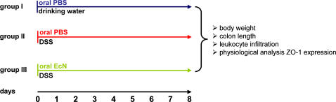
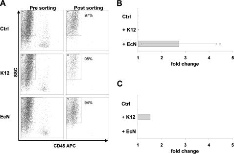
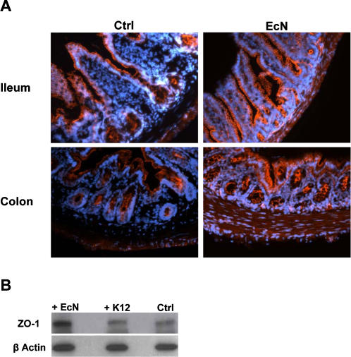
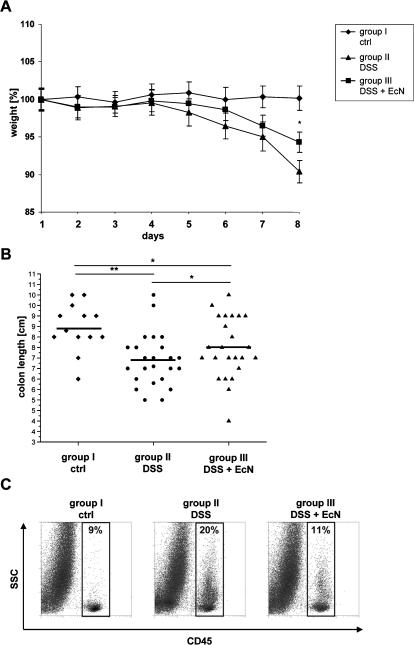
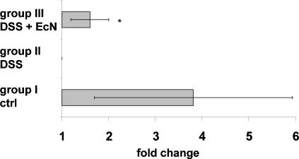
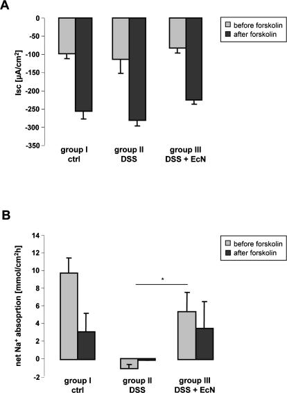
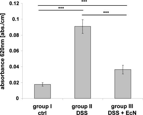
Methodology and principal findings: To study the in vivo effect of probiotic Escherichia coli Nissle 1917 (EcN) on the stabilization of the intestinal barrier under healthy conditions, germfree mice were colonized with EcN or K12 E. coli strain MG1655. IECs were isolated and analyzed for gene and protein expression of the tight junction molecules ZO-1 and ZO-2. Then, in order to analyze beneficial effects of EcN under inflammatory conditions, the probiotic was orally administered to BALB/c mice with acute dextran sodium sulfate (DSS) induced colitis. Colonization of gnotobiotic mice with EcN resulted in an up-regulation of ZO-1 in IECs at both mRNA and protein levels. EcN administration to DSS-treated mice reduced the loss of body weight and colon shortening. In addition, infiltration of the colon with leukocytes was ameliorated in EcN inoculated mice. Acute DSS colitis did not result in an anion secretory defect, but abrogated the sodium absorptive function of the mucosa. Additionally, intestinal barrier function was severely affected as evidenced by a strong increase in the mucosal uptake of Evans blue in vivo. Concomitant administration of EcN to DSS treated animals resulted in a significant protection against intestinal barrier dysfunction and IECs isolated from these mice exhibited a more pronounced expression of ZO-1.

Conclusion and significance: This study convincingly demonstrates that probiotic EcN is able to mediate up-regulation of ZO-1 expression in murine IECs and confer protection from the DSS colitis-associated increase in mucosal permeability to luminal substances.


Conflict of interest statement

Competing Interests: The authors have declared that no competing interests exist.

Figures

Figure 1
Figure 1. Experimental design of the DSS colitis model.
Acute colitis was induced by administration of 4–6% DSS to drinking water (DSS). (A) Group I was orally treated with PBS twice daily. Group II was given 4–6% DSS in drinking water and orally treated with PBS twice daily. Group III received 1.5–2×10⁸ CFU EcN two times a day by oral application in combination with 4–6% DSS in drinking water (DSS+EcN).
Figure 2
Figure 2. Isolation of IECs from gnotobiotic mice colonized with EcN or E. coli MG1655 and ZO-1 expression analysis.
Gnotobiotic BALB/c mice were colonized with E. coli MG1655 (K12) or EcN for 6 days, respectively. Application of PBS was used as control. (A) Whole intestinal cell populations were isolated from gnotobiotic mice treated either with PBS, K12 or EcN. For sorting of a pure intestinal epithelial cell population, cells were labeled with anti-CD45 antibody to exclude hematopoietic cells and further distinguished by cell granularity and size (SSC) (Pre sorting). IECs were FACS-sorted by negative selection. Re-analysis was performed to determine the purity of sorted IECs (Post sorting). (B) Quantitative ZO-1 mRNA expression in IECs. Relative mRNA amounts were normalized with respect to expression levels of IECs from control mice (fold change = 1). Data are presented as mean of three independent experiments (n = 3/group). (C) Quantitative ZO-2 mRNA expression in IECs. Relative mRNA amounts were normalized with respect to expression levels of IECs from control mice (fold change = 1). Data are presented as mean of three independent experiments (n = 3/group). *p<0.05 EcN vs. Ctrl (relative expression values).
Figure 3
Figure 3. ZO-1 protein expression in ileum and colon of gnotobiotic mice.
(A) Immunofluorescence staining of tissue sections from gnotobiotic control mice and mice colonized with EcN for 6 days with a fluorescent anti-ZO-1 antibody (orange). Original magnification×20. (B) Protein expression of ZO-1 in IECs. Western blot analysis of FACS sorted IECs from colonized mice was performed using anti-ZO-1 antibody. Anti-β actin antibody was used as internal control, binding to the corresponding protein with a molecular weight of 42 kDa. The ZO-1 antibody detects endogenous ZO-1 protein, displayed as a band at ∼210 kDa.
Figure 4
Figure 4. Administration of EcN in DSS induced colitis.
(A) Disease severity was measured daily and is expressed in terms of body weight loss. (group II: n = 23, group III: n = 25, group I: n = 17) *p<0.05 group III vs. group II. (B) Reduction of colon length. Measurement of colon length [cm] after preparation. (group II: n = 24, group III: n = 25, group I: n = 13) *p<0.05 group III vs. group II or group III vs. group I **p<0.01 group II vs. group I. (C) Infiltration of hematopoietic cells into the colon. Whole intestinal cell populations were labeled with anti-CD45 APC antibody and measured by FACS analysis (n = 6/group).
Figure 5
Figure 5. Increased ZO-1 mRNA expression in mice treated with DSS and EcN.
IECs were isolated from indicated mice and relative levels of ZO-1 mRNA were normalized with respect to the expression level of IECs from DSS treated mice (fold change = 1). Data are presented as mean of four independent experiments (n = 3/group). *p<0.05 group III vs. group II (relative expression values).
Figure 6
Figure 6. Secretory and absorptive function of isolated colonic mucosa from DSS-treated mice, with and without EcN administration, and healthy controls.
(A) Basal and forskolin-stimulated Isc in DSS treated (group II n = 7), DSS+EcN (group III n = 12) and control animals (group I n = 10). The different numbers resulted from the fact that considerably more colonic segments from the DSS treated mice were so friable that they ruptured before measurements could be taken. (B) Basal and post-forskolin net Na+ flux rates in the three different groups. Whereas Na+ absorption was completely abolished in group II, active Na+ absorption was only partially inhibited in group III. Only in the control group I could an inhibition of Na+ absorption by forskolin be observed, indicating normal regulation (n = 6/group). *p<0.05
Figure 7
Figure 7. Permeability to Evans Blue in the colon of DSS treated (group II), DSS+EcN (group III) treated mice and healthy controls (group I).
The graph shows a significant increase in Evans Blue uptake into the colonic mucosa of group II mice and a strong reduction in Evans Blue uptake in group III mice to almost normal values (n = 6/group). ***p<0.001

References

    1. Farquhar MG, Palade GE. Junctional complexes in various epithelia. J Cell Biol. 1963;17:375–412. - PMC - PubMed
    1. Kapembwa MS, Fleming SC, Sewankambo N, Serwadda D, Lucas S, et al. Altered small-intestinal permeability associated with diarrhoea in human-immunodeficiency-virus-infected Caucasian and African subjects. Clin Sci (Lond) 1991;81:327–334. - PubMed
    1. Rene E, Marche C, Regnier B, Saimot AG, Vilde JL, et al. Intestinal infections in patients with acquired immunodeficiency syndrome. A prospective study in 132 patients. Dig Dis Sci. 1989;34:773–780. - PubMed
    1. Smith PD, Lane HC, Gill VJ, Manischewitz JF, Quinnan GV, et al. Intestinal infections in patients with the acquired immunodeficiency syndrome (AIDS). Etiology and response to therapy. Ann Intern Med. 1988;108:328–333. - PubMed
    1. Stevenson BR, Siliciano JD, Mooseker MS, Goodenough DA. Identification of ZO-1: a high molecular weight polypeptide associated with the tight junction (zonula occludens) in a variety of epithelia. J Cell Biol. 1986;103:755–766. - PMC - PubMed

Publication types