. 2007 Dec;5(12):e325.
doi: 10.1371/journal.pbio.0050325.

The foxa2 gene controls the birth and spontaneous degeneration of dopamine neurons in old age

Affiliations

The foxa2 gene controls the birth and spontaneous degeneration of dopamine neurons in old age

Raja Kittappa et al. PLoS Biol. 2007 Dec.

Abstract

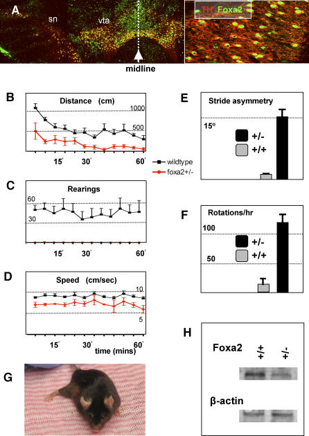
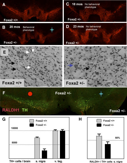
Parkinson disease affects more than 1% of the population over 60 y old. The dominant models for Parkinson disease are based on the use of chemical toxins to kill dopamine neurons, but do not address the risk factors that normally increase with age. Forkhead transcription factors are critical regulators of survival and longevity. The forkhead transcription factor, foxa2, is specifically expressed in adult dopamine neurons and their precursors in the medial floor plate. Gain- and loss-of-function experiments show this gene, foxa2, is required to generate dopamine neurons during fetal development and from embryonic stem cells. Mice carrying only one copy of the foxa2 gene show abnormalities in motor behavior in old age and an associated progressive loss of dopamine neurons. Manipulating forkhead function may regulate both the birth of dopamine neurons and their spontaneous death, two major goals of regenerative medicine.


Conflict of interest statement

Competing interests. The authors have declared that no competing interests exist.

Figures

Figure 1
Figure 1. Floor Plate Origin of Dopamine Neurons
(A and B) Initial differentiation of dopamine neurons is in the medial domain of the foxa2-expressing floor plate. Low (A) and high (B) magnification images of E12.5 midbrain stained for FOXA2 (red) and tyrosine hydroxylase (green). Note that the newly born dopamine neurons continue to express foxa2. (C) Schematic of the ventral progenitor domains of the embryonic mesencephalon. Dopamine precursors (DA) occupy the ventral-most position in the mesencephalon, the medial floor plate (MFP), whereas the lateral floor plate (LFP) is comprised of oculomotor neuron precursors (MN). V3 precursors are on either side of the floor plate, in the ventral basal plate (BP). V3 precursors are not the most-ventral neuronal progenitor in the midbrain, as they are in the spinal cord, because mesencephalic floor plate cells undergo neurogenesis. The expression of LMX1b, PHOX2a, and NKX2.2 expression are shown in Figures S2 and S3. NKX6.1 expression in the midbrain is shown in Figure S3. (D) SHH (red) and LMX1b (green) are coexpressed in the floor plate of the embryonic midbrain. (E) ß-galactosidase staining in the E15.5 midbrain of a shh-cregfp:: ROSA26 mouse fetus. In this mouse, ß-galactosidase is expressed in the shh-expressing cells of the floor plate and their derivatives. Inset contains a low-power image of a coronal section of the midbrain showing that the blue ß-galactosidase signals is restricted to the most ventral region. (F–H) Immunohistochemical localization in (F) and (G) shows that both TH (green) and ß-galactosidase (red) are expressed in the same cells, demonstrating a floor plate origin for dopamine neurons. (G) Inset: enlargement of boxed cells showing colocalization of TH and ß-galactosidase at the single-cell level. (H) Another enlarged field contains numerous neurons coexpressing TH and ß-galactosidase.
Figure 2
Figure 2. The In Vitro Role of foxa2 and SHH in Cell Fate Specification in the Ventral Mesencephalon
(A) Effects of Shh on E8.5 mesencephalic precursors in vitro. In the absence of exogenous SHH, many LMX1b+ precursors (36.59 ± 6.10%) were observed. NKX2.2+ (19.83 ± 4.39%), NKX6.1+ (30.47 ± 6.72%), and PAX7+ precursors (16.33 ± 5.02%) were present. In the presence of 500 ng/ml of SHH protein, the proportion of NKX2.2+ (41.84 ± 5.84%) and NKX6.1+ (56.62 ± 7.04%) cells increased, whereas, the proportion of PAX7+ cells diminished (6.89 ± 2.54%). Despite these effects on other cell types, the proportion of LMX1b+ dopamine precursors was stable (32.48 ± 5.56%). Higher concentrations of SHH would be expected to further ventralize the midbrain precursors at the expense of more dorsal fates. In the presence of 1 μg/ml of SHH protein, there was a further increase in NKX2.2+ and NKX6.1+ cells (nkx2.2+, 52.75 ± 6.34%; nkx6.1+, 69.12 ± 8.73%) and a greater reduction of PAX7+ cells (2.24 ± 0.81%). At this high concentration of SHH, the percentage of dopamine precursors again remained unchanged (33.04 ± 6.61%). (B) SHH has a proliferative effect on mesencephalic precursors in cell culture. BrdU was added to day 3 cultures of mesencephalic precursors, grown in different concentrations of SHH protein. BrdU was added for 1 h before fixation and staining. The percentage of cells incorporating BrdU increased with the concentration of SHH in the culture. (C) Wild-type mesencephalic explants differentiate to generate a large number of neurons (Tuj1, green) and a significant proportion of these neurons express tyrosine hydroxylase (TH, red). (D) Mesencephalic explants from foxa2−/− embryos also generate many neurons, but TH expression is absent. (E) foxa2−/− explants similarly do not yield islet-1–expressing motor neurons although the differentiation of GATA3- and LIM1/2-positive neurons born outside of the floor plate is unaffected. The developmental expression pattern of these proteins can be seen in Figure S3. (F) Overexpression of foxa2 in cultured E10.5 mesencephalic explants results in an increase in TH-expressing dopamine neurons. (G) Doxycycline induction of foxa2 in differentiating F4 mouse embryonic stem cells significantly increases the number of resulting dopamine neurons. (H) Inhibition of SHH signaling by cyclopamine suppresses dopaminergic differentiation of ES cells. The induction of foxa2 by doxycycline overcomes cyclopamine suppression of dopamine neuron differentiation.
Figure 3
Figure 3. foxa2 and the Adult Midbrain
(A) Coronal section through adult mouse midbrain stained showing that foxa2 is expressed in dopamine neurons of the SN and VTA of the adult mouse midbrain. ([A], inset) Enlargement of cells in (A) showing colocalization of TH and FOXA2 at the single cell level. (B–E) Analyses of motor behavior in mutant and wild-type mice. (B) Horizontal movement of mutant (red) and wild-type (blue) mice in the activity monitor. (C) Vertical movement (rearing) of mutant (red) and wild-type (blue) mice in the activity monitor. Note that mutant mice do not rear at all. (D) Speed of mutant (red) mice is compromised relative to wild-type (blue) mice. (E) Footprint analysis was used to quantitate the angle of spinal curvature in mutant mice and age-matched wild-type mice. (F) Amphetamine significantly induces rotational movement in foxa2+/− as compared to wild-type controls. (G) Image of 18-mo-old foxa2+/− mouse with a rigid right rear limb. (H) Western analysis of matched 1-mm sections of ventral midbrain shows a reduction of FOXA2 protein in foxa2+/− mice as compared to wild-type controls. As a loading control for total protein, beta-actin staining is shown below.
Figure 4
Figure 4. Dopamine Neuron Loss in foxa2+/− Mice
(A and B) Dopamine neurons in a 24-mo-old wild-type mouse (A) and a foxa2+/− littermate (B). The mouse in (B) possessed a kinked posture and the other late-onset motor phenotypes. A significant loss of nigral dopamine neurons is easily observed on one side of the brain marked by a blue star in the foxa2+/− mouse. (C and D) TH staining of the the ventral midbrain in older foxa2+/− animals without late onset behavioral abnormalities. Note that the dopaminergic system resembles the wild-type condition seen in (A). (E) Nissl staining of wild-type and foxa2+/− mutant mice. Representative glia and neurons are highlighted with white and black arrows, respectively. Note the significant loss of normal neuronal morphologies in the mutant animal. Occasionally, a dysmorphic cell, highlighted here with a purple arrow, can be seen in the mutant, but not the wild-type brain. (F) Ventral tier dopamine neurons, stained for RALDH1 (red), are selectively lost in foxa2+/− animals. In this animal, nigral dopamine neurons are almost completely lost on the left side (marked by the blue star), whereas the right side (marked by the red sun) is much less affected (note that these are coronal sections, presented as if the animal were facing the viewer, so that the left side of the animal is on the viewer's right side. (G) Quantitation of dopamine neurons in the SN and VTA of wild-type (n = 3) and foxa2+/− (n = 3) mice. The number of dopamine neurons in the mutant SN (2,765.0 ± 750.5) is substantially smaller than in the control SN (7,344.3 ± 197.9). In contrast, the number of the dopamine neurons in the VTA is roughly the same in wild-type (9,922.0 ± 375.7) and mutant mice (10,064.7 ± 355.4). (H) The percentage of RALDH1-expressing neurons in the SN is reduced in foxa2+/− mutants as compared to wild-type controls.

Similar articles

Cited by

References

    1. Lewis SJ, Dove A, Robbins TW, Barker R, Owen AM. Cognitive impairments in early Parkinson's disease are accompanied by reductions in activity in frontostriatal neural circuitry. J. Neurosci. 2003;23:6351–6. - PMC - PubMed
    1. Lange KW, Paul GM, Robbins TW, Marsden CD. L-dopa and frontal cognitive function in Parkinson's disease. Adv Neurol. 1993;60:475–478. - PubMed
    1. de Rijk MC, Launer LJ, Berger K, Breteler MM, Dartigues JF, et al. Prevalence of Parkinson's Disease in Europe: a collaborative study of population-based cohorts. Neurology. 2000;54:S21–S23. - PubMed
    1. Altman J, Bayer S.A. Development of the brain stem in the rat. V. Thymidine-radiographic study of the time of origin of neurons in the midbrain tegmentum. J Comp Neurol. 1981;198:677–716. - PubMed
    1. Studer L, Tabar V, McKay RD. Transplantation of expanded mesencephalic precursors leads to recovery in parkinsonian rats. Nat Neurosci. 1998;1:290–295. - PubMed

Publication types

MeSH terms