Case Reports
. 2007 Dec 12;49(1):37.
doi: 10.1186/1751-0147-49-37.

Strongyloides stercoralis infection in a Finnish kennel

Affiliations
Case Reports

Strongyloides stercoralis infection in a Finnish kennel

Kati J Dillard et al. Acta Vet Scand. .

Abstract

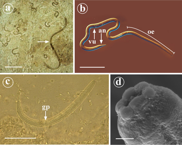
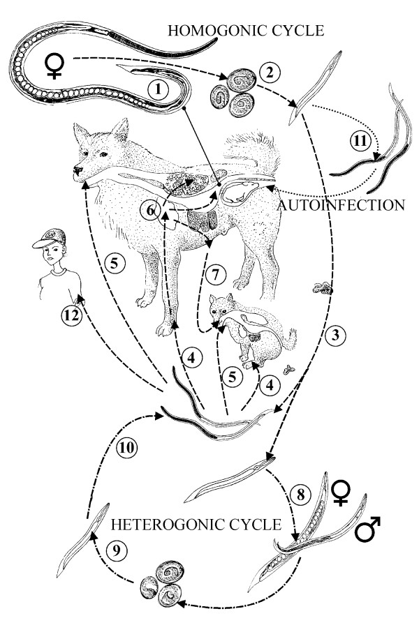
Background: Intestinal threadworm Strongyloides stercoralis is a parasite of dog, cat and primates that occurs worldwide being most prevalent in tropical and subtropical countries. The adult parasitic worm is about 2 mm long and slender. It possesses both parasitic and free-living lifecycles. The parasitic worms are females. Strongyloides stercoralis infects the host via percutaneous, peroral or transmammary transmission in addition to autoinfection. Clinical disease varies from inapparent to severe enteritis and pneumonia. The diagnosis is based on demonstration of larvae in fresh faeces, which is best made by Baermann technique.

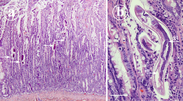
Case presentation: Strongyloides stercoralis infection was diagnosed in autopsy in a 10-week-old puppy born and raised in a Finnish kennel. Prior to its sudden death, the puppy had suffered from gastrointestinal disturbance for three weeks. Subsequent sampling of the dogs in the kennel revealed that three adult dogs in the kennel were also infected.

Conclusion: The present case shows that S. stercoralis can complete its life cycle and cause disease in dogs also in Northern Europe. Infection can be maintained also in a temperate climate and may become a chronic problem in a kennel environment. Infection may be underdiagnosed as Baermann technique is not routinely performed in small animal practice.


Figures

Figure 1
Figure 1
Parasitological findings detected in 10-week-old Yorkshire terrier puppy suffering from Strongyloides stercoralis infection. Figure 1a: In the intestinal scrapings of the duodenum numerous adult small female nematodes (arrow), larvae and ova were found. Scale bar = 200 μm. Figure 1b: Adult parasitic female possesses long cylindrical oesophagus (oe) that occupies the anterior third of the body. Vulva (vu) and anus (an) are located in the posterior third of the body and the tail is narrowly tapered. Scale bar = 200 μm. Figure 1c: First stage larva of S. stercoralis. Genital primordium (gp) is very prominent. Scale bar = 50 μm. Figure 1d: The anterior end of a parasitic female S. stercoralis as observed under SEM. Hexagonal oral opening is surrounded by six well-defined lips. Scale bar = 2 μm.
Figure 2
Figure 2
Histopathology as seen in the duodenal mucosa of 10-week-old Yorkshire terrier puppy suffering from S. stercoralis infection. Figure 2a: A micrograph to show numerous S. stercoralis larvae and ova (arrows) within the intestinal mucosa. In the mucosa there is moderate diffuse infiltration of lymphocytes and plasma cells. Haematoxylin-eosin stained histological section, scale bar = 50 μm. Figure 2b: A close-up micrograph with female S. stercoralis. The small size of the parasite, the relatively large intestine (in) and paired genital tract (gt) are readily seen in this longitudinally sectioned female. Haematoxylin-eosin stained histological section, scale bar = 50 μm.
Figure 3
Figure 3
Life cycle of S. stercoralis. All adult parasites are partenogenetic females (1) that reside in the crypts of small intestine. Their embryonated eggs (2) hatch in the crypts or the intestinal lumen. The L1 rhabditiform larvae are voided in faeces (3) and give a rise either to L3 infective filariform larvae (L3i) or develop to free living adult nematodes depending on environmental conditions. The L3i develops no further if it does not gain access to new host. The L3i enters the dog percutaneously (4) perorally(5). The larvae migrate to the small intestine and molt first to L4 and then tothe parthenogenic adult female(6). The puppies can be infected via milk if the bitch has migrating L3 larvae (7). Transplacental infection does not occur. This parasitic life cycle of S. stercoralis is referred as homogonic life cycle. If environmental conditions are optimal, an alternative route of life cycle (heterogonic life cycle) can take place. Non-infective rhabditiform larvae develop to free-living adult male and female worms (8) that produce eggs. Non-infectious rhabditiform larvae (9) hatching from the eggs will develop to L3i (10). During passage through the host intestinal tract, rhabditiform larvae may rapidly undergo molts into L3i. These larvae can penetrate through the wall of large intestine or perianal skin of the host resulting in migration ending in the small intestine (11). The process is called autoinfection and it is favoured especially in neonatal or immunocompromized hosts. The canine strains of S. stercoralis have been known to infect humans (12). The life cycle was drawn based on the information obtained from following references: [2,4,16–18].

References

    1. Roberts LS, Janovy J. Nematodes: Rhabditida, pioneering parasites. In: Gerald D. Schmidt, Larry S, editor. Roberts' Foundations of parasitology. New York, USA, Mc Graw-Hill; 2005. pp. 411–416.
    1. Georgi JR, Georgi ME. Strongyloides. In: Georgi JR, Georgi ME, editor. Canine Clinical Parasitology. Malvern, PA, USA, Lea & Febiger; 1992. pp. 160–165.
    1. Grove DI. Human Strogyloidiasis. In: Baker JR, Muller R, Dollison R, editor. Advances in Parasitology. Vol. 38. Academic Press; 1996. pp. 251–309. - PubMed
    1. Nolan TJ. Canine Strongyloidiasis. 2001. http://www.ivis.org/advances/Parasit_Bowman/nolan_strongyloidiasis/chapt... Accessed May 30, 2007.
    1. Bowman DD. Georgi's parasitology for veterinarians. USA, W. B. Saunders Company; 2003. pp. 197–200.

Publication types

LinkOut - more resources