. 2007;8(12):R264.
doi: 10.1186/gb-2007-8-12-r264.

Evolution of allostery in the cyclic nucleotide binding module

Affiliations

Evolution of allostery in the cyclic nucleotide binding module

Natarajan Kannan et al. Genome Biol. 2007.

Abstract

Background: The cyclic nucleotide binding (CNB) domain regulates signaling pathways in both eukaryotes and prokaryotes. In this study, we analyze the evolutionary information embedded in genomic sequences to explore the diversity of signaling through the CNB domain and also how the CNB domain elicits a cellular response upon binding to cAMP.

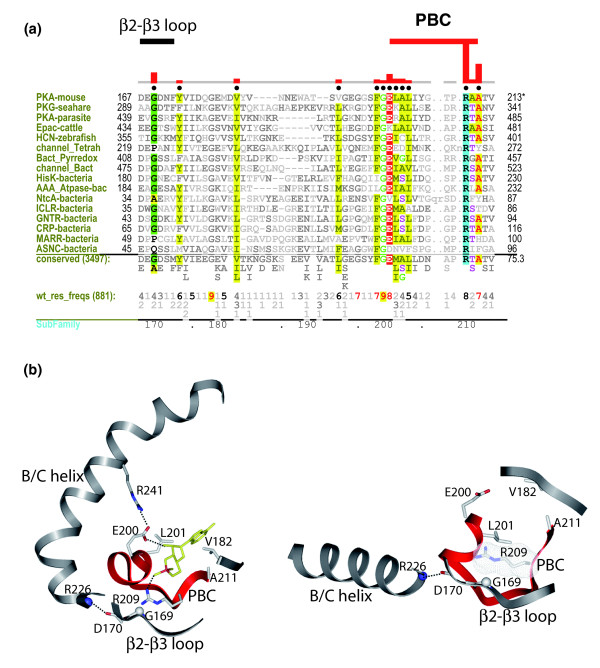
Results: Identification and classification of CNB domains in Global Ocean Sampling and other protein sequences reveals that they typically are fused to a wide variety of functional domains. CNB domains have undergone major sequence variation during evolution. In particular, the sequence motif that anchors the cAMP phosphate (termed the PBC motif) is strikingly different in some families. This variation may contribute to ligand specificity inasmuch as members of the prokaryotic cooA family, for example, harbor a CNB domain that contains a non-canonical PBC motif and that binds a heme ligand in the cAMP binding pocket. Statistical comparison of the functional constraints imposed on the canonical and non-canonical PBC containing sequences reveals that a key arginine, which coordinates with the cAMP phosphate, has co-evolved with a glycine in a distal beta2-beta3 loop that allosterically couples cAMP binding to distal regulatory sites.

Conclusion: Our analysis suggests that CNB domains have evolved as a scaffold to sense a wide variety of second messenger signals. Based on sequence, structural and biochemical data, we propose a mechanism for allosteric regulation by CNB domains.


Figures

Figure 1
Figure 1
Classification and domain organization of CNB domain containing families. (a) Phylogenetic tree of the 30 identified families. Eukaryotic branches are shown in dark teal, while the prokaryotic branches are shaded in gold. Novel families in bacteria are indicated by red dots. Families that have a non-canonical PBC are indicated by blue dots. (b) Domain organization of known and novel CNB domain containing proteins in eukaryotes and prokaryotes.
Figure 2
Figure 2
Conserved features of the CNB domain. A contrast hierarchical alignment showing conserved residues/motifs shared by the entire superfamily. The histograms above the alignments plot the strength of the selective constraints imposed at each position. Secondary structure is indicated directly above the aligned sequences with β-strands indicated by their number designations (that is, 1-7 correspond to the β1-β7 strands, respectively) and helices by their letter designations. The leftmost column of each alignment shows the sequences used in the display alignment. See Materials and methods for sequence identifiers. The background alignment of all CNB domain containing sequences are shown indirectly via the consensus patterns and corresponding weighted residue frequencies ('wt_res_freqs') below the display alignment. (Such sequence weighting adjusts for overrepresented families in the alignment.) The residue frequencies are indicated in integer tenths where, for example, a '5' indicates that the corresponding residue directly above it occurs in 50-60% of the weighted sequences. Biochemically similar residues are colored similarly with the intensity of the highlighting proportional to how strikingly foreground residues contrast with background residues.
Figure 3
Figure 3
The structural location of the conserved glycines in the PKA regulatory subunit R1alpha (PDB: 1RGS). The alpha subdomain is shown in light gray and the beta subdomain is shown in dark grey. The glycines are shown in spheres representation.
Figure 4
Figure 4
Core conserved residues shared by the entire superfamily and the conformational changes associated with the helical subdomain. (a) cAMP bound structure of the PKA regulatory subunit R1alpha (PDB: 1RGS). (b) Catalytic subunit (C-subunit) bound structure of R1alpha (PDB: 2QCS). The alpha subdomain is shown in yellow and the beta subdomain is shown in white. The PBC region is colored in red. The hydrophobic residues are shown in sticks and surface representation, and the glycine residues are shown in CPK representation. The core conserved residues are colored in gold.
Figure 5
Figure 5
Sequence variation within the PBC and ligand specificity. (a) A schematic representation of the PBC showing the secondary structures and the consensus motif. (b) Families that contain a canonical and non-canonical PBC motif. Sequence alignment of the PBC region showing conserved and variable positions. Conserved residues are highlighted and Arg209 position is indicated by a black box. (c-f) The conformation of the PBC region in: the PKA regulatory subunit (PDB: 1RGS) (c); PDZ_GEF (PDB: 2D93) (d); cooA (PDB: 1FT9) (e); CprK (PDB: 2H6B) (f).
Figure 6
Figure 6
Sequence features that distinguish the canonical and non-canonical PBC containing sequences. (a) A contrast hierarchical alignment (see Figure 2 legend) showing residues (indicated by black dots above alignment) that distinguish the canonical PBC containing sequences from the non-canonical ones. Biochemically similar residues are colored similarly with the intensity of the highlighting proportional to how strikingly foreground residues contrast with background residues. (b) The allosteric link between the PBC and β2-β3 loop is shown using the cAMP bound and cAMP-free structures of the PKA regulatory subunit.

References

    1. Berman HM, Ten Eyck LF, Goodsell DS, Haste NM, Kornev A, Taylor SS. The cAMP binding domain: an ancient signaling module. Proc Natl Acad Sci USA. 2005;102:45–50. doi: 10.1073/pnas.0408579102. - DOI - PMC - PubMed
    1. Anantharaman V, Koonin EV, Aravind L. Regulatory potential, phyletic distribution and evolution of ancient, intracellular small-molecule-binding domains. J Mol Biol. 2001;307:1271–1292. doi: 10.1006/jmbi.2001.4508. - DOI - PubMed
    1. Gill GN, Garren LD. Role of the receptor in the mechanism of action of adenosine 3':5'-cyclic monophosphate. Proc Natl Acad Sci USA. 1971;68:786–790. doi: 10.1073/pnas.68.4.786. - DOI - PMC - PubMed
    1. Taylor SS, Buechler JA, Yonemoto W. cAMP-dependent protein kinase: framework for a diverse family of regulatory enzymes. Annu Rev Biochem. 1990;59:971–1005. doi: 10.1146/annurev.bi.59.070190.004543. - DOI - PubMed
    1. de Rooij J, Zwartkruis FJ, Verheijen MH, Cool RH, Nijman SM, Wittinghofer A, Bos JL. Epac is a Rap1 guanine-nucleotide-exchange factor directly activated by cyclic AMP. Nature. 1998;396:474–477. doi: 10.1038/24884. - DOI - PubMed

Publication types

LinkOut - more resources