. 2008 Feb;126(2):189-201.
doi: 10.1016/j.clim.2007.10.004.

A novel subset of memory B cells is enriched in autoreactivity and correlates with adverse outcomes in SLE

Affiliations

A novel subset of memory B cells is enriched in autoreactivity and correlates with adverse outcomes in SLE

Matilda W Nicholas et al. Clin Immunol. 2008 Feb.

Abstract

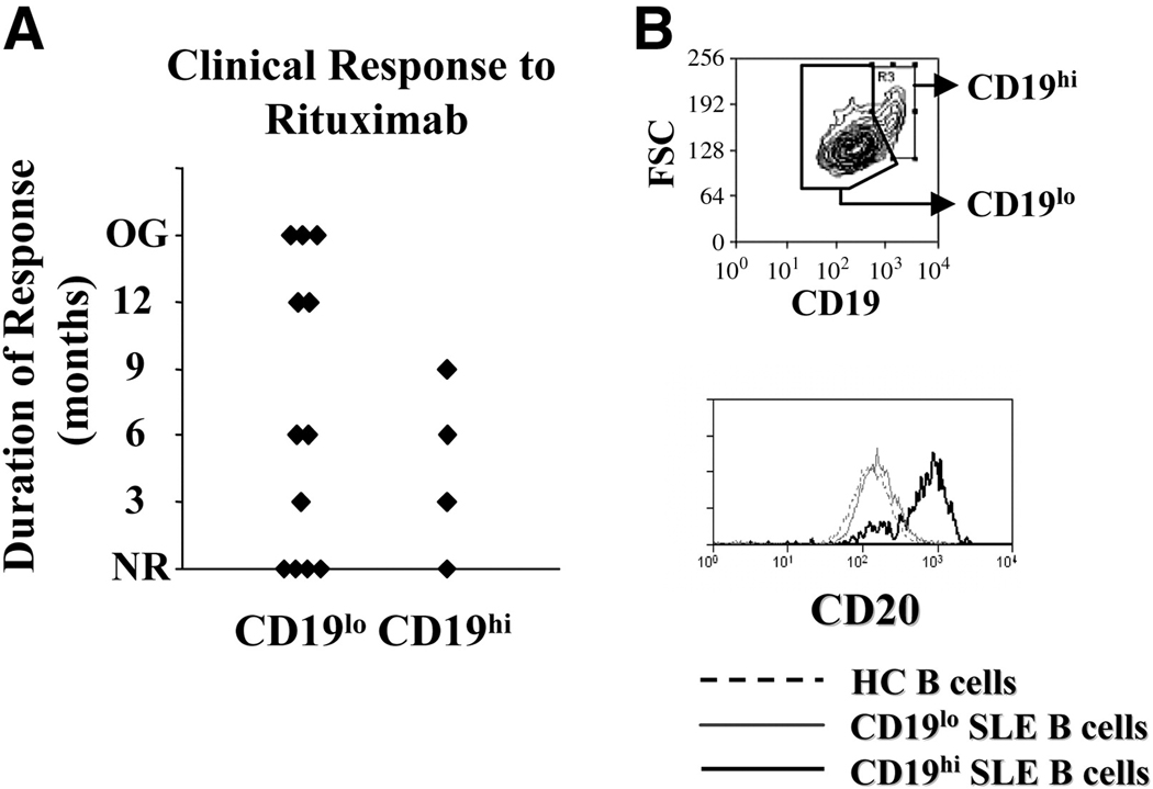
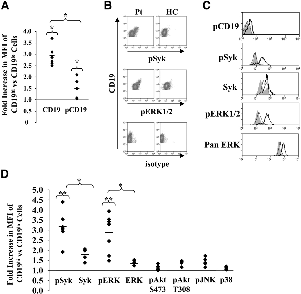
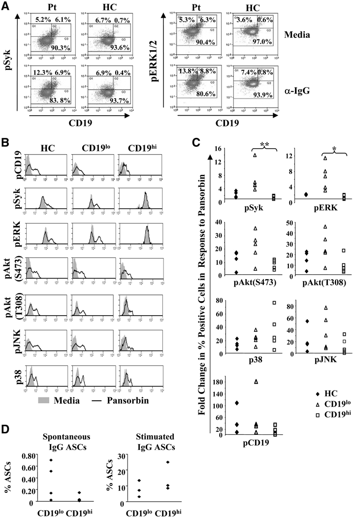
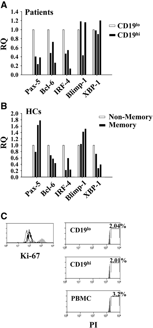
We previously reported that some systemic lupus erythematosus (SLE) patients have a population of circulating memory B cells with >2-fold higher levels of CD19. We show here that the presence of CD19(hi) B cells correlates with long-term adverse outcomes. These B cells do not appear anergic, as they exhibit high basal levels of phosphorylated Syk and ERK1/2, signal transduce in response to BCR crosslinking, and can become plasma cells (PCs) in vitro. Autoreactive anti-Smith (Sm) B cells are enriched in this population and the degree of enrichment correlates with the log of the serum anti-Sm titer, arguing that they undergo clonal expansion before PC differentiation. PC differentiation may occur at sites of inflammation, as CD19(hi) B cells have elevated CXCR3 levels and chemotax in response to its ligand CXCL9. Thus, CD19(hi) B cells are precursors to anti-self PCs, and identify an SLE patient subset likely to experience poor clinical outcomes.


Figures

Fig. 1
Fig. 1. CD19hi patients are poor responders to rituximab treatment
A, Clinical response of CD19hi SLE patients and CD19lo SLE patients to rituximab treatment. Response was defined as an improvement in the SLAM or SLEDAI score of at least 3 points and duration of response defined as the time point at which these disease activity indexes increased by 3 points or an increase in steroid dosage was required. NR=no response; OG=ongoing response. B, Representative histograms showing gating for CD19hi and CD19lo B cells from an SLE patient. Only CD19+ PB B cells are included in this gate. The bottom histogram shows CD20 expression by CD19hi and CD19lo B cells and healthy control B cells (all CD19+ B cells). Results are representative of 3 patients.
Fig. 2
Fig. 2. CD19hi B cells are enriched for autoreactivity, and the degree of autoreactivity in these cells correlates exponentially with serum autoantibody titers
A, CD19hi and CD19lo B cells were sorted (using gates illustrated in Fig. 1B) from four patients with differing anti-Sm titers, and ELISpot used to determine the number of anti-Sm IgG ASCs in each group with and without stimulation by pansorbin, anti-CD40, IL-10 and IL-2. B, Anti-Sm ASCs per million CD19hi or CD19lo B cells in each of four patients. Patient 1 did not have detectable serum anti-Sm titer, and no anti-Sm ASCs were seen in either cell group. C, Anti-Sm ASCs per million cells in each group vs. log10[anti-Sm serum titer], showing an exponential relationship between the number of anti-Sm ASCs in the CD19hi B cell group and the serum titer of anti-Sm in the corresponding patient.
Fig. 3
Fig. 3. Basal phosphorylative state of CD19hi cells
PB cells were permeabilized and stained using antibodies recognizing the indicated molecules. Gating for CD19hi and CD19lo B cells is shown in Fig. 1B. A, Fold increase in MFIs of CD19 or phosphorylated CD19 (pCD19) in CD19hi vs CD19lo cells. B, Representative histograms of PB cells from a CD19hi patient and healthy control (HC) stained for CD19 and pSyk, pERK1/2, or an isotype control. Gating is on all CD19+ B cells. C, Representative single parameter histograms showing staining for phosphorylated CD19 and phosphorylated or total Syk and ERK1/2 in PB B cells from a healthy control (shaded) and CD19lo (thin line) and CD19hi (thick line) B cells from the same patient. Gating is on CD19hi and CD19lo B cells as shown in Fig. 1B. D, Fold increases in MFI for the labeled signaling molecules in CD19lo vs. CD19hi cells from the same patient. A and D, Each dot represents a different patient and/or day. At least four different patients are included for each signaling molecule. * p≤0.05; **p≤0.001.
Fig. 4
Fig. 4. CD19hi B cells respond to BCR crosslinking
A, Representative histograms of PB B cells from a CD19hi patient and a healthy control after incubation with media alone or with anti-IgG for 10 min. Histograms are gated on CD19+ PB B cells and the percentages give are of B cells falling within each indicated gate. B, Representative single parameter histograms for each of the indicated phosphorylated molecules in PB B cells from a healthy control and CD19lo and CD19hi B cells from a patient after incubation with media (shaded) or pansorbin (line) for 10 min. Gating is illustrated in Fig. 1B. C, Fold change in the percentage of cells falling into the positive gate for each of the phosphorylated signaling molecules tested in cells incubated with media or pansorbin in a healthy control or in CD19lo and CD19hi B cells from the same patient. Each point represents a different individual. D, CD19hi and CD19lo B cells (see Fig. 2A) were sorted and IgG antibody secreting cells (ASCs) determined after incubation with either media alone or with pansorbin, anti-CD40, IL-10 and IL-2 for 6 days. Each dot represents a different patient. * p≤0.05; **p≤0.001.
Fig. 5
Fig. 5. CD19hi B cells have altered transcription factor expression
A and B, Transcription factor expression in indicated sorted B cell groups as determined by RQ RT-PCR. Each bar represents a different individual. Gating for cell sorting is shown in Fig. 1B. A, Relative expression in CD19hi B cells as compared to CD19lo B cells from the same patient. B, Relative expression in CD19+CD38IgD memory B cells as compared to all other CD19+ B cells from the same healthy control. C, Representative histograms for Ki-67 staining in non-lymphoid PBMCs (shaded) and CD19lo (thin line) or CD19hi (thick line) patient PB B cells and for PI staining of indicated subsets. Percentages indicate cells falling into drawn gate.
Fig 6
Fig 6. CXCR expression in patient CD19hi and healthy control memory B cells
A and B, CXCR expression in indicated sorted B cell groups as determined by RQ RT-PCR. Each bar represents a different individual. A, Relative expression in CD19hi B cells as compared to CD19lo B cells from the same patient. B, Representative histogram of CD19+ B cells from a healthy control showing gating of memory B cells (circled; CD38-, IgD-). The non-memory B cells used were gated as all CD19+ B cells that fell outside this gate. Bars represent relative transcript expression in CD19+CD38IgD memory B cells as compared to all other CD19+ B cells from the same healthy control. C, Surface CXCR3 expression determined by flow cytometry on healthy control B cells (shaded) or in CD19lo (thin line) or CD19hi (thick line) B cells from the same patient. Histogram is representative of three patients. D, Chemotaxis of CD19hi B cells in response to the CXCR3 ligand CXCL9, the CXCR4 ligand CXCL12, or media alone. Migration was determined by collection, staining, and flow cytometry of cells which migrated into lower wells after 90 minutes. No chemotaxis was observed in response to CXCL12 and media alone by cells from patient 2.

References

    1. Hahn BH. Antibodies to DNA. N Engl J Med. 1998;338:1359–1368. - PubMed
    1. Su W, Madaio MP. Recent advances in the pathogenesis of lupus nephritis: autoantibodies and B cells. Semin Nephrol. 2003;23:564–568. - PubMed
    1. Harris DP, Haynes L, Sayles PC, Duso DK, Eaton SM, Lepak NM, Johnson LL, Swain SL, Lund FE. Reciprocal regulation of polarized cytokine production by effector B and T cells. Nat Immunol. 2000;1:475–482. - PubMed
    1. Bijl M, Horst G, Limburg PC, Kallenberg CG. Expression of costimulatory molecules on peripheral blood lymphocytes of patients with systemic lupus erythematosus. Ann Rheum Dis. 2001;60:523–526. - PMC - PubMed
    1. Chan OT, Hannum LG, Haberman AM, Madaio MP, Shlomchik MJ. A novel mouse with B cells but lacking serum antibody reveals an antibody-independent role for B cells in murine lupus. J Exp Med. 1999;189:1639–1648. - PMC - PubMed

Publication types

MeSH terms