Comparative Study
. 2008 Aug 6;5(25):855-63.
doi: 10.1098/rsif.2007.1274.

Selective activation of mechanosensitive ion channels using magnetic particles

Affiliations
Comparative Study

Selective activation of mechanosensitive ion channels using magnetic particles

Steven Hughes et al. J R Soc Interface. .

Abstract

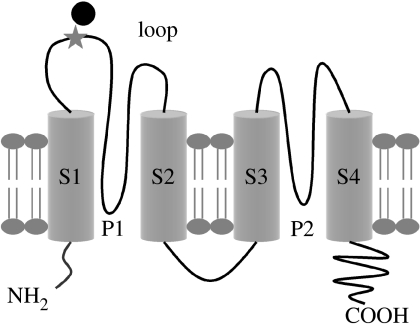
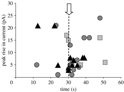
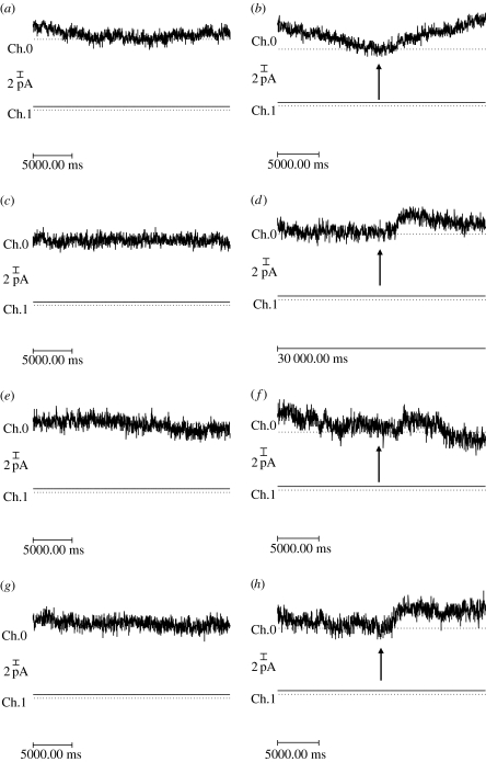
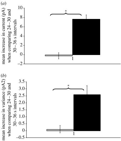
This study reports the preliminary development of a novel magnetic particle-based technique that permits the application of highly localized mechanical forces directly to specific regions of an ion-channel structure. We demonstrate that this approach can be used to directly and selectively activate a mechanosensitive ion channel of interest, namely TREK-1. It is shown that manipulation of particles targeted against the extended extracellular loop region of TREK-1 leads to changes in whole-cell currents consistent with changes in TREK-1 activity. Responses were absent when particles were coated with RGD (Arg-Gly-Asp) peptide or when magnetic fields were applied in the absence of magnetic particles. It is concluded that changes in whole-cell current are the result of direct force application to the extracellular loop region of TREK-1 and thus these results implicate this region of the channel structure in mechano-gating. It is hypothesized that the extended loop region of TREK-1 may act as a tension spring that acts to regulate sensitivity to mechanical forces, in a nature similar to that described for MscL. The development of a technique that permits the direct manipulation of mechanosensitive ion channels in real time without the need for pharmacological drugs has huge potential benefits not only for basic biological research of ion-channel gating mechanisms, but also potentially as a tool for the treatment of human diseases caused by ion-channel dysfunction.


Figures

Figure 1
Figure 1
Schematic of the ‘6.His.loop’ mTREK-1 clone indicating the position of the inserted 6.His repeat (star) and attached magnetic particles (black circle; image not shown to scale). Adapted from Patel et al. (2001).
Figure 2
Figure 2
(a) Representative whole-cell ‘ramp’ and ‘steps’ recordings (−100 to +100 mV) from (i,ii) wild-type TREK-1 and (iii,iv) 6.His.loop.TREK-1 transfected COS-7 cells. Ch.0=membrane current (Im) and Ch.1=membrane voltage (Vm). (b) Comparison of the mean whole-cell current recorded at +80 mV from 6.His.loop.TREK-1 transfected cells in the presence (black bar, n=25) and the absence (grey bar, n=11) of attached ‘His-targeted’ magnetic particles at 48 hours post-transfection. Data shown are pooled from experiments using a range of different magnetic particles (range 250 nm to 2.7 μm).
Figure 3
Figure 3
(a) Cell-attached recording of 6.His.loop.TREK-1 in response to negative pipette pressure applied to the cell membrane via the patch pipette during a+50 mV voltage step. Pressure applied as indicated by arrow. (b) Whole-cell ‘ramp’ recordings from a 6.His.loop.TREK-1 transfected COS-7 cell before and after the addition of LPC (100 μM) to the recording bath. Time to onset of response was 100 s and time to peak increase was 290 s. Ch.0=Im and Ch.1=Vm. Traces are recorded in the absence of magnetic particles.
Figure 4
Figure 4
The percentage of ‘no magnet’, ‘static magnet’ and ‘1 Hz magnet’ traces recorded from 6.His.loop.TREK-1 transfected COS-7 cells exhibiting changes in outward current classified as events. (a) Data shown are pooled from all experiments (including replicants) using Ni–NTA and anti-His antibody coated magnetic particles of all sizes. Cells patched 24–72 hours post-transfection. n refers to the total number of traces and includes replicants from individual cells. Light grey bars: no magnet (+80 mV: n=57;+50 mV: n=54), black bars: static magnet (+80 mV: n=43;+50 mV: n=40) and dark grey bars: 1 Hz magnet (+80 mV: n=54; +50 mV: n=36). (b) Response pooled by particle size and holding potential.
Figure 5
Figure 5
The time of onset and peak rise in outward current for all ‘events’ detected during no magnet (squares), static magnet (circles) and 1 Hz magnet (triangles) stimulation experiments. Data points shown are pooled from all experiments performed with anti-His and Ni–NTA-coated particles ranging in size from 250 nm to 2.7 μm. Onset of magnetic stimulation is indicated by the dashed line.
Figure 6
Figure 6
A series of traces showing (a,c,e,g) ‘no magnet’ whole-cell currents and whole-cell currents recorded in response to the application of (b,d,f) static and (h) 1 Hz time-varying magnetic fields (80 mT) recorded from the same 6.His.loop.TREK-1 transfected cell with 6×1 μm anti-His antibody coated particles attached to its surface. For all traces, whole-cell current is recorded for a period of 60 s following a voltage step to +80 mV from a holding potential of 0 mV, where magnetic field exposure was commenced at t=30 s (as indicated by arrows). Images shown are magnified regions of the whole-cell traces. Records were performed in the order shown (i.e. a,b,c, etc). Ch.0=Im and Ch.1=Vm.
Figure 7
Figure 7
Graphs showing mean change in (a) current and (b) variance observed when comparing 24–30 (before magnet) and 30–36 (after magnet) seconds time intervals for static magnet (black bar) and no magnet (grey bar) experiments (n=3) performed on a 6.His.loop.TREK-1 transfected COS-7 cell with anti-His antibody coated magnetic particles attached (traces are shown in figure 6). Asterisk denotes p<0.05.

References

    1. Ajouz B, Berrier C, Besnard M, Martinac B, Ghazi A. Contributions of the different extramembranous domains of the mechanosensitive ion channel MscL to its response to membrane tension. J. Biol. Chem. 2000;275:1015–1022. doi: 10.1074/jbc.275.2.1015. - DOI - PubMed
    1. Alloui A, et al. TREK-1, a K+ channel involved in polymodal pain perception. EMBO J. 2006;25:2368–2376. doi: 10.1038/sj.emboj.7601116. - DOI - PMC - PubMed
    1. Browe D.M, Baumgarten C.M. Stretch of beta 1 integrin activates an outwardly rectifying chloride current via FAK and Src in rabbit ventricular myocytes. J. Gen. Physiol. 2003;122:689–702. doi: 10.1085/jgp.200308899. - DOI - PMC - PubMed
    1. Cartmell S, El Haj A.J. Mechanical bioreactors for tissue engineering. In: Chaudhuri J.B, Rubeai M.A, editors. Bioreactors for tissue engineering, principles, design and operation. Springer; Berlin, Germany: 2005. pp. 193–208. ch. 8.
    1. Chemin J, Patel A.J, Duprat F, Lauritzen I, Lazdunski M, Honore E. A phospholipid sensor controls mechanogating of the K(+) channel TREK-1. EMBO J. 2005a;24:44–53. doi: 10.1038/sj.emboj.7600494. - DOI - PMC - PubMed

Publication types

MeSH terms

Substances