. 2007 Dec 18;104(51):20308-13.
doi: 10.1073/pnas.0710517105. Epub 2007 Dec 12.

Dual-color superresolution imaging of genetically expressed probes within individual adhesion complexes

Affiliations

Dual-color superresolution imaging of genetically expressed probes within individual adhesion complexes

Hari Shroff et al. Proc Natl Acad Sci U S A. .

Erratum in

  • Proc Natl Acad Sci U S A. 2008 Sep 30;105(39):15220

Abstract

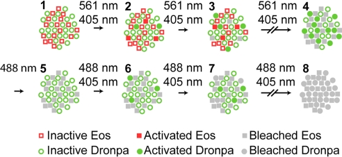
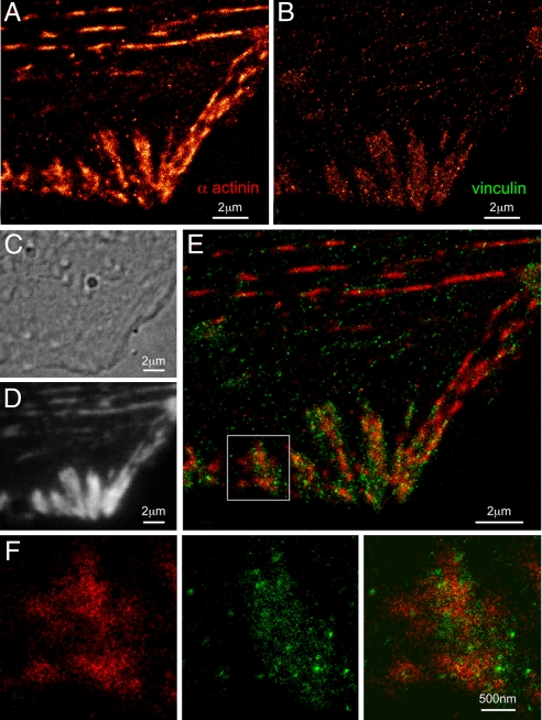
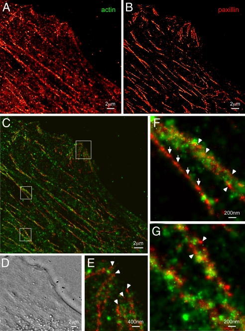
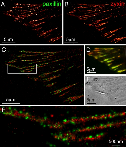
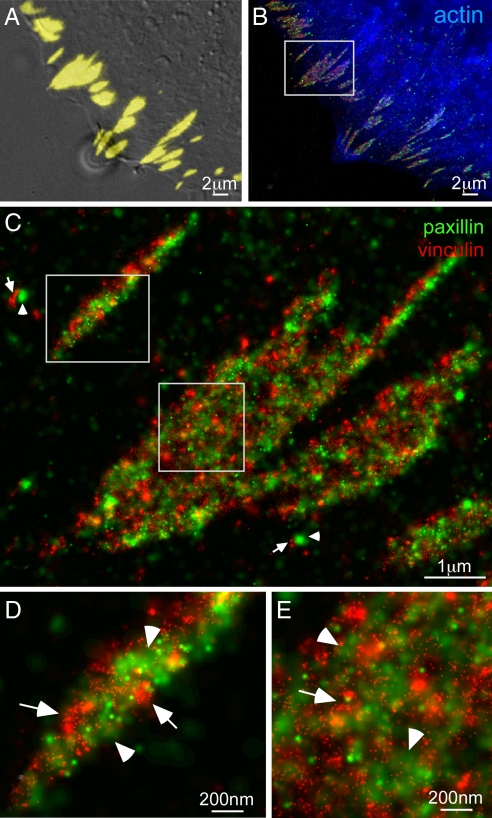
Accurate determination of the relative positions of proteins within localized regions of the cell is essential for understanding their biological function. Although fluorescent fusion proteins are targeted with molecular precision, the position of these genetically expressed reporters is usually known only to the resolution of conventional optics ( approximately 200 nm). Here, we report the use of two-color photoactivated localization microscopy (PALM) to determine the ultrastructural relationship between different proteins fused to spectrally distinct photoactivatable fluorescent proteins (PA-FPs). The nonperturbative incorporation of these endogenous tags facilitates an imaging resolution in whole, fixed cells of approximately 20-30 nm at acquisition times of 5-30 min. We apply the technique to image different pairs of proteins assembled in adhesion complexes, the central attachment points between the cytoskeleton and the substrate in migrating cells. For several pairs, we find that proteins that seem colocalized when viewed by conventional optics are resolved as distinct interlocking nano-aggregates when imaged via PALM. The simplicity, minimal invasiveness, resolution, and speed of the technique all suggest its potential to directly visualize molecular interactions within cellular structures at the nanometer scale.


Conflict of interest statement

The authors declare no conflict of interest.

Figures

Fig. 1.
Fig. 1.
Protocol for dual-label superresolution imaging by PALM. A specimen initially expressing inactive EosFP and Dronpa molecules (step 1) is exposed to a 405-nm activation light and a 561-nm Eos-excitation light (steps 2 and 3) until all EosFP molecules are detected, localized, and bleached (step 4). The many active Dronpa molecules that then exist (step 4) are deactivated by using an intense 488-nm light (step 5). Both 405-nm and 488-nm light are then applied (steps 6 and 7) to serially activate, detect, localize, and eventually bleach (step 8) all remaining Dronpa molecules. PALM images encompassing 105 to 106 molecules are thereby acquired, typically in 5–30 min each.
Fig. 2.
Fig. 2.
Nanostructural organization of cytoskeletal α-actinin and adhesion complex localized vinculin in an HFF-1 cell. (A) PALM image of Dronpa-tagged α-actinin. (B) PALM image of tdEos-tagged vinculin. (C) DIC image revealing morphology. (D) TIRF image of combined tdEos and Dronpa emission. (E) Dual-color PALM overlay of α-actinin (red) and vinculin (green). (F) α-Actinin, vinculin, and overlaid PALM images within the single adhesion shown in the box in E. E and F reveal that α-actinin and vinculin only partially colocalize within each adhesion, with α-actinin existing in large patches emanating from stress fibers and vinculin coalescing in small, dense clusters scattered across each adhesion.
Fig. 3.
Fig. 3.
Nanostructural organization of cytoskeletal actin and the adhesion protein paxillin in an HFF-1 cell. (A) PALM image of Dronpa-tagged actin. (B) PALM image of tdEos-tagged paxillin. (C) Dual-color PALM overlay of actin (green) and paxillin (red). (D) DIC image in the same region. Paxillin assembles in fibrillar-like adhesions that run parallel to ventral actin fibers and cluster near the cell periphery to form larger adhesion complexes (topmost box in C, shown expanded in E). Very little direct overlap is observed between actin and paxillin, although actin bundles densely cluster around some (arrowheads in E–G) but not all (full arrows in F) fibrillar paxillin adhesions.
Fig. 4.
Fig. 4.
Dual-color PALM using PS-CFP2 as an alternative second label. (A) PALM image of tdEos-tagged paxillin. (B) PALM image of PS-CFP2-tagged zyxin. (C) Dual-color PALM overlay of paxillin (green) and zyxin (red). (D) Diffraction-limited, summed molecule, dual-color TIRF image (8). (E) DIC image. The two adhesion proteins seem colocalized in D, but are revealed in C and the boxed region shown at higher magnification in F to have very little overlap, with paxillin tightly clustered in separate nano-domains.
Fig. 5.
Fig. 5.
Triple-label imaging with combined techniques. (A) Overlaid DIC and TIRF (yellow) images of paxillin and vinculin coexpressed in an HFF-1 cell. (B) Diffraction-limited epi-fluorescence image of mCerulean-tagged actin (blue) overlaid with PALM images of Dronpa-tagged paxillin (green) and tdEos-tagged vinculin (red) shows adhesion complexes at the periphery of the cell aligned with the termini of actin bundles. An expanded view (C) of the boxed region in B reveals parallel arrays of interwoven paxillin and vinculin aggregates along the length of each AC, as well as possibly nascent adhesion complexes consisting of adjacent paxillin (arrowheads) and vinculin aggregates (arrows). Further magnified views (D and E) of the boxed regions in C indicate other examples of adjacent aggregates of either paxillin (arrowheads) or vinculin (arrows) within larger adhesions.

References

    1. Giepmans BNG, Adams SR, Ellisman MH, Tsien RY. The fluorescent toolbox for assessing protein location and function. Science. 2006;312:217–224. - PubMed
    1. Hwang J, Gheber LA, Margolis L, Edidin M. Domains in cell plasma membranes investigated by near-field scanning optical microscopy. Biophys J. 1998;74:2184–2190. - PMC - PubMed
    1. Enderle T, Ha T, Ogletree DF, Chemla DS, Magowan C, Weiss S. Membrane specific mapping and colocalization of malarial and host skeletal proteins in the Plasmodium falciparum infected erythrocyte by dual-color near-field scanning optical microscopy. Proc Natl Acad Sci USA. 1997;94:520–525. - PMC - PubMed
    1. Donnert G, Keller J, Wurm CA, Rizzoli SO, Westphal V, Schönle A, Jahn R, Jakobs S, Eggeling C, Hell SW. Two-color far-field fluorescence nanoscopy. Biophys J. 2007;92:L67–L69. - PMC - PubMed
    1. Gaietta G, Deerinck TJ, Adams SR, Bouwer J, Tour O, Laird DW, Sosinsky GE, Tsien RY, Ellisman MH. Multicolor and electron microscopic imaging of connexin trafficking. Science. 2002;296:503–507. - PubMed

Publication types