. 2007 Dec 18;104(51):20466-71.
doi: 10.1073/pnas.0710335105. Epub 2007 Dec 12.

Diet-induced obesity in mice causes changes in immune responses and bone loss manifested by bacterial challenge

Affiliations

Diet-induced obesity in mice causes changes in immune responses and bone loss manifested by bacterial challenge

Salomon Amar et al. Proc Natl Acad Sci U S A. .

Abstract

Obesity has been suggested to be associated with an increased susceptibility to bacterial infection. However, few studies have examined the effect of obesity on the immune response to bacterial infections. In the present study, we investigated the effect of obesity on innate immune responses to Porphyromonas gingivalis infection, an infection strongly associated with periodontitis. Mice with diet-induced obesity (DIO) and lean control C57BL/6 mice were infected orally or systemically with P. gingivalis, and periodontal pathology and systemic immune responses were examined postinfection. After oral infection with P. gingivalis, mice with DIO had a significantly higher level of alveolar bone loss than the lean controls. Oral microbial sampling disclosed higher levels of P. gingivalis in mice with DIO vs. lean mice during and after infection. Furthermore, animals with DIO exposed to oral infection or systemic inoculation of live P. gingivalis developed a blunted inflammatory response with reduced expression of TNF-alpha, IL-6, and serum amyloid A (SAA) at all time points compared with lean mice. Finally, peritoneal macrophages harvested from mice with DIO and exposed to P. gingivalis exhibited reduced levels of proinflammatory cytokines compared with lean mice and when exposed to P. gingivalis LPS treatment had a significantly reduced recruitment of NF-kappaB to both TNF-alpha and IL-10 promoters 30 min after exposure. These data indicate that obesity interferes with the ability of the immune system to appropriately respond to P. gingivalis infection and suggest that this immune dysregulation participates in the increased alveolar bone loss after bacterial infection observed in mice with DIO.


Conflict of interest statement

The authors declare no conflict of interest.

Figures
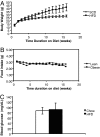
Fig. 1.
Fig. 1.
DIO mouse model used in the study. Four-week-old male Jackson Laboratory C57BL/6 mice were fed either with SCD or HFD for 16 weeks. Body weight was measured weekly (A). Mean food intake (B) and serum glucose levels (C) were measured before experiments.
Fig. 2.
Fig. 2.
P. gingivalis-induced alveolar bone loss in experimental periodontitis in both lean mice and mice with DIO. Mice from both groups were exposed to P. gingivalis-soaked or broth-soaked ligatures. After four challenges with bacteria or broth alone, mice were euthanized, bone tissue was prepared, and bone measurements were performed by morphometric analysis (A). Distance from the cemental enamel junction (CEJ) to the alveolar bone crest (ABC) was measured in millimeters, and the data presented are means ± SEM (B) (n = 5 for each group). *, P = 0.0007.
Fig. 3.
Fig. 3.
TNF-α levels in sera from mice with experimental periodontitis induced by P. gingivalis. Groups of DIO and lean mice were infected with P. gingivalis in the form of ligature insertions at the second molar on day 0, and ligatures were replaced on days 3, 5, and 7. Blood samples were collected before ligature insertion at days 0, 3, 5, 7, 10, and 12. TNF-α levels in the sera were determined by ELISA (BioSource). *, P < 0.05.
Fig. 4.
Fig. 4.
Serum TNF-α (A), IL-6 (B), and SAA (C) levels in response to infection with P. gingivalis strain A7436 in lean and mice with DIO by the intravenous route. Mice were infected with P. gingivalis (2.0 × 10⁹ in 50 μl) or vehicle by tail vein injection on days 1, 3, and 5. TNF-α, IL-6, and SAA concentrations in serum were measured by ELISA (means ± SEM, n = 8). *, P < 0.05.
Fig. 5.
Fig. 5.
Global cytokine profile in response to P. gingivalis stimulation in vitro. Macrophages from lean mice and mice with DIO were cultured and exposed to live P. gingivalis for 15 min and 4 h (MOI = 25:1). Cells treated with PBS were used as controls. Culture supernatants were analyzed by a Bio-Plex cytokine array. Cytokines induced after 15-min and 4-h treatment and their average concentrations (pg/ml) (means ± SD; n = 3) are shown. *, P < 0.01 between lean and DIO. Cytokines that did not change significantly between lean and obese were the following: eotaxin, IFN-γ, IL-2, IL-3, IL-4, IL-5, IL-9, IL-10, IL-12p70, IL-13, and IL-17.
Fig. 6.
Fig. 6.
Effects of DIO on macrophage response to P. gingivalis LPS. Mouse macrophages isolated from lean mice and mice with DIO were challenged with PBS or P. gingivalis LPS for 4 h and 24 h. TNF-α concentrations in cell culture supernatants were measured by ELISA (n = 3). *, P < 0.05; **, P < 0.001.
Fig. 7.
Fig. 7.
Differential gene expression between macrophages from mice with DIO and lean mice after P. gingivalis LPS challenge. Data show the relative gene expression level in DIO over that from lean mice. Negative values signify gene expression lower in DIO mouse macrophages compared with lean counterparts.
Fig. 8.
Fig. 8.
Early and transient recruitment of NF-κB to the TNF-α and IL-10 promoters. Peritoneal macrophages from lean mice and mice with DIO were treated with P. gingivalis LPS (10 μg/ml) for the indicated times, and ChIP assays were performed with an anti-p65 affinity-purified rabbit polyclonal Ab. p65-precipitated DNA was analyzed by quantitative real-time PCR with promoter-specific primers amplifying the TNF-α and IL-10 promoters. The results are expressed as means ± SD; n = 4 for each time point. *, P < 0.05; **, P < 0.001.

References

    1. Dasanayake AP, et al. Preterm low birth weight and periodontal disease among African Americans. Dent Clin North Am. 2003;47:115–125. x–xi. - PubMed
    1. Nishimura F, et al. Periodontal infection and dyslipidemia in type 2 diabetics: Association with increased HMG-CoA reductase expression. Horm Metab Res. 2006;38:530–535. - PubMed
    1. Campus G, Salem A, Uzzau S, Baldoni E, Tonolo G. Diabetes and periodontal disease: A case-control study. J Periodontol. 2005;76:418–425. - PubMed
    1. Chi H, Messas E, Levine RA, Graves DT, Amar S. Interleukin-1 receptor signaling mediates atherosclerosis associated with bacterial exposure and/or a high-fat diet in a murine apolipoprotein E heterozygote model: Pharmacotherapeutic implications. Circulation. 2004;110:1678–1685. - PubMed
    1. Chiang CY, Kyritsis G, Graves DT, Amar S. Interleukin-1 and tumor necrosis factor activities partially account for calvarial bone resorption induced by local injection of lipopolysaccharide. Infect Immun. 1999;67:4231–4236. - PMC - PubMed

Publication types

MeSH terms