. 2007 Dec 26;104(52):20908-13.
doi: 10.1073/pnas.0710054104. Epub 2007 Dec 11.

Prion strain discrimination in cell culture: the cell panel assay

Affiliations

Prion strain discrimination in cell culture: the cell panel assay

Sukhvir P Mahal et al. Proc Natl Acad Sci U S A. .

Abstract

Prions are thought to consist mainly or entirely of misfolded PrP, a constitutively expressed host protein. Prions associated with the same PrP sequence may occur in the form of different strains; the strain phenotype is believed to be encoded by the conformation of the PrP. Some cell lines can be persistently infected by prions and, interestingly, show preference for certain strains. We report that a cloned murine neuroblastoma cell population, N2a-PK1, is highly heterogeneous in regard to its susceptibility to RML and 22L prions. Remarkably, sibling subclones may show very different relative susceptibilities to the two strains, indicating that the responses can vary independently. We have assembled four cell lines, N2a-PK1, N2a-R33, LD9 and CAD5, which show widely different responses to prion strains RML, 22L, 301C, and Me7, into a panel that allows their discrimination in vitro within 2 weeks, using the standard scrapie cell assay (SSCA).


Conflict of interest statement

The authors declare no conflict of interest.

Figures
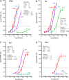
Fig. 1.
Fig. 1.
Responsiveness of PK1, CAD5, LD9, and R33 cells to various prion strain preparations. The cells indicated in the figure (PK1, blue; CAD5, red; LD9, violet; R33, green) were infected with prion preparations and subjected to the SSCA. The cells were infected with a serial 1:3 dilution of 0.1% homogenates of brains infected with RML (a), 22L (b), Me7 (c), and 301C (d) prions. Response Index300,3 (RI300,3 or RI for short) of a cell line for a prion strain is defined as the reciprocal of the dilution required to yield 300 scrapie-positive cells per 20,000 cells after the third split. The characteristic metric adopted in this article is the ratio of the RI of a cell line relative to that given by LD9 cells.
Fig. 2.
Fig. 2.
Responsiveness of PK1 subclones to 22L and RML prions. (a) Sixteen clones derived from the PK1 population (CAB clones, filled circles), as well as PK1 (open triangle) and R33 (open circle) were expanded to confluence in wells of a 96-well plate, and 20,000 cells of each were challenged with 10−5 RML and 22L-infected brain homogenates, respectively. The SSCA was performed for three splits as described. The spot numbers are plotted against each other. Two of the PK1 subclones are marginally more responsive to both prion strains than the original PK1 population whereas the remainder lost responsiveness to both strains to varying degrees. (b) One clone from a (encircled) was used to generate a second generation of 552 subclones, which were challenged with a 10−4 dilution of RML or 22L-infected brain homogenate. The responses of the parental clone are depicted with a star. (c) Third-generation subclones (264) were isolated from one clone from b (encircled) and also challenged with 10−4 dilutions of RML or 22L brain homogenate. Again, the responses of the parental clone from b are shown as a star in c. The axis labels in c refer to all graphs.
Fig. 3.
Fig. 3.
Correlation between response indexes and total PrP or doubling time. The data are from Table 1. (a and b) Total PrP versus the RIs for RML and 22L. (c and d) Doubling times versus the RIs for RML and 22L, respectively, of R33, PK1, and various PK1-derived CAB clones. The lowercase letter next to the points refers to the clones listed in Table 1.

Comment in

Similar articles

Cited by

References

    1. Prusiner SB. Annu Rev Microbiol. 1989;43:345–374. - PubMed
    1. Tremblay P, Ball HL, Kaneko K, Groth D, Hegde RS, Cohen FE, DeArmond SJ, Prusiner SB, Safar JG. J Virol. 2004;78:2088–2099. - PMC - PubMed
    1. Tzaban S, Friedlander G, Schonberger O, Horonchik L, Yedidia Y, Shaked G, Gabizon R, Taraboulos A. Biochemistry. 2002;41:12868–12875. - PubMed
    1. Safar J, Wille H, Itri V, Groth D, Serban H, Torchia M, Cohen FE, Prusiner SB. Nat Med. 1998;4:1157–1165. - PubMed
    1. Pastrana MA, Sajnani G, Onisko B, Castilla J, Morales R, Soto C, Requena JR. Biochemistry. 2006;45:15710–15717. - PubMed

Publication types