

A systematic interaction map of validated kinase inhibitors with Ser/Thr kinases
- PMID: 18077363
- PMCID: PMC2154464
- DOI: 10.1073/pnas.0708800104
A systematic interaction map of validated kinase inhibitors with Ser/Thr kinases
Abstract
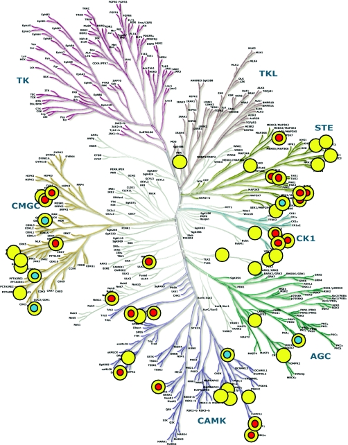
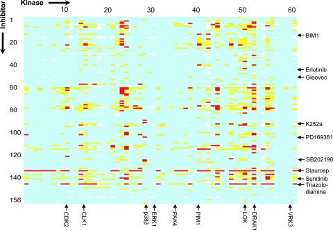
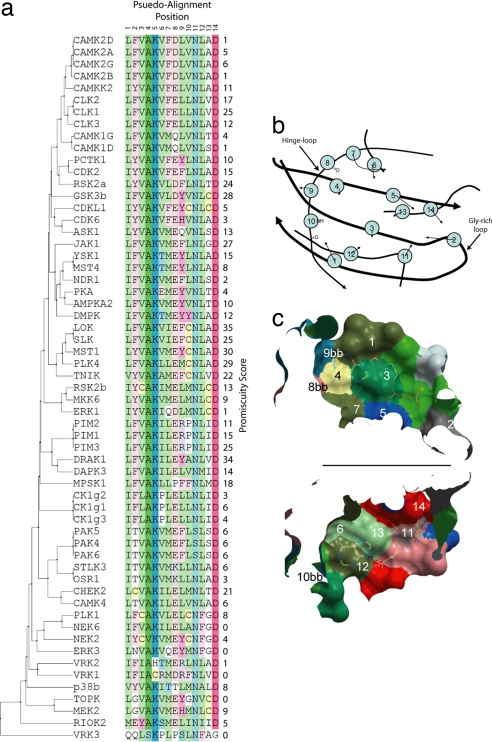
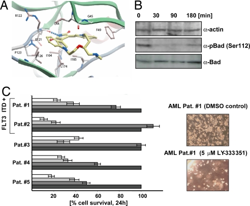
Protein kinases play a pivotal role in cell signaling, and dysregulation of many kinases has been linked to disease development. A large number of kinase inhibitors are therefore currently under investigation in clinical trials, and so far seven inhibitors have been approved as anti-cancer drugs. In addition, kinase inhibitors are widely used as specific probes to study cell signaling, but systematic studies describing selectivity of these reagents across a panel of diverse kinases are largely lacking. Here we evaluated the specificity of 156 validated kinase inhibitors, including inhibitors used in clinical trials, against 60 human Ser/Thr kinases using a thermal stability shift assay. Our analysis revealed many unexpected cross-reactivities for inhibitors thought to be specific for certain targets. We also found that certain combinations of active-site residues in the ATP-binding site correlated with the detected ligand promiscuity and that some kinases are highly sensitive to inhibition using diverse chemotypes, suggesting them as preferred intervention points. Our results uncovered also inhibitor cross-reactivities that may lead to alternate clinical applications. For example, LY333'531, a PKCbeta inhibitor currently in phase III clinical trials, efficiently inhibited PIM1 kinase in our screen, a suggested target for treatment of leukemia. We determined the binding mode of this inhibitor by x-ray crystallography and in addition showed that LY333'531 induced cell death and significantly suppressed growth of leukemic cells from acute myeloid leukemia patients.
Conflict of interest statement
The authors declare no conflict of interest.
Figures
References
-
- Manning G, Whyte DB, Martinez R, Hunter T, Sudarsanam S. Science. 2002;298:1912–1934. - PubMed
-
- Cohen P. Nat Rev Drug Discovery. 2002;1:309–315. - PubMed
-
- Imming P, Sinning C, Meyer A. Nat Rev Drug Discovery. 2006;5:821–834. - PubMed
-
- Vieth M, Sutherland JJ, Robertson DH, Campbell RM. Drug Discovery Today. 2005;10:839–846. - PubMed
-
- Ohno R. Int J Clin Oncol. 2006;11:176–183. - PubMed
Publication types
MeSH terms
Substances
Associated data
- Actions
Grants and funding
LinkOut - more resources
Full Text Sources
Other Literature Sources
Chemical Information
Molecular Biology Databases
