

Deletion of the BH3-only protein puma protects motoneurons from ER stress-induced apoptosis and delays motoneuron loss in ALS mice
- PMID: 18077368
- PMCID: PMC2154478
- DOI: 10.1073/pnas.0707906105
Deletion of the BH3-only protein puma protects motoneurons from ER stress-induced apoptosis and delays motoneuron loss in ALS mice
Erratum in
-
Correction for Kieran et al., Deletion of the BH3-only protein puma protects motoneurons from ER stress-induced apoptosis and delays motoneuron loss in ALS mice.Proc Natl Acad Sci U S A. 2021 Aug 17;118(33):e2112400118. doi: 10.1073/pnas.2112400118. Proc Natl Acad Sci U S A. 2021. PMID: 34380742 Free PMC article. No abstract available.
Abstract
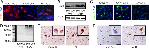
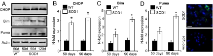
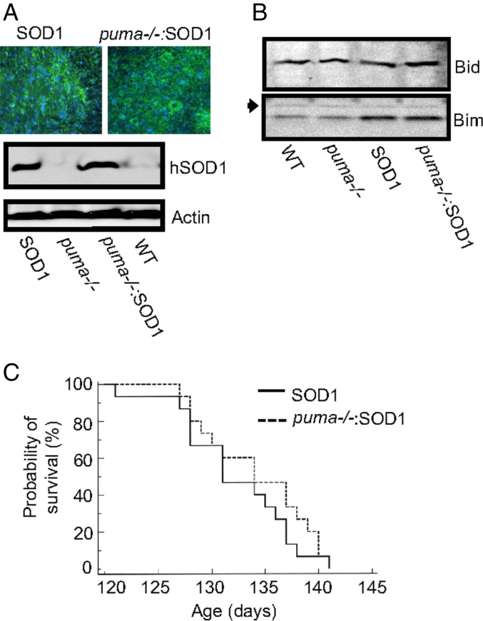
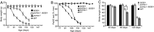
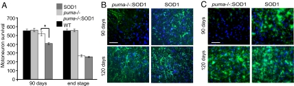
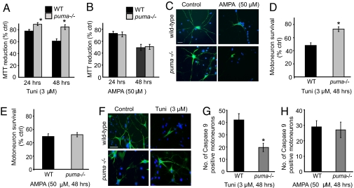
BH3-only proteins couple diverse stress signals to the evolutionarily conserved mitochondrial apoptosis pathway. Previously, we reported that the activation of the BH3-only protein p53-up-regulated mediator of apoptosis (Puma) was necessary and sufficient for endoplasmic reticulum (ER) stress- and proteasome inhibition-induced apoptosis in neuroblastoma and other cancer cells. Defects in protein quality control have also been suggested to be a key event in ALS, a fatal neurodegenerative condition characterized by motoneuron degeneration. Using the SOD1(G93A) mouse model as well as human post mortem samples from ALS patients, we show evidence for increased ER stress and defects in protein degradation in motoneurons during disease progression. Before symptom onset, we detected a significant up-regulation of Puma in motoneurons of SOD1(G93A) mice. Genetic deletion of puma significantly improved motoneuron survival and delayed disease onset and motor dysfunction in SOD1(G93A) mice. However, it had no significant effect on lifespan, suggesting that other ER stress-related cell-death proteins or other factors, such as excitotoxicity, necrosis, or inflammatory injury, may contribute at later disease stages. Indeed, further experiments using cultured motoneurons revealed that genetic deletion of puma protected motoneurons against ER stress-induced apoptosis but showed no effect against excitotoxic injury. These findings demonstrate that a single BH3-only protein, the ER stress-associated protein Puma, plays an important role during the early stages of chronic neurodegeneration in vivo.
Conflict of interest statement
The authors declare no conflict of interest.
Figures
References
-
- Cleveland DW, Rothstein JD. Nat Rev Neurosci. 2001;2:806–819. - PubMed
-
- Rosen DR, Siddique T, Patterson D, Figlewicz DA, Sapp P, Hentati A, Donaldson D, Goto J, O'Regan JP, Deng HX, et al. Nature. 1993;362:59–62. - PubMed
-
- Gurney ME, Pu H, Chiu AY, Dal Canto MC, Polchow CY, Alexander DD, Caliendo J, Hentati A, Kwon YW, Deng HX, et al. Science. 1994;264:1772–1775. - PubMed
-
- Shibata N, Hirano A, Kobayashi M, Siddique T, Deng HX, Hung WY, Kato T, Asayama K. J Neuropathol Exp Neurol. 1996;55:481–490. - PubMed
-
- Bruijn LI, Becher MW, Lee MK, Anderson KL, Jenkins NA, Copeland NG, Sisodia SS, Rothstein JD, Borchelt DR, Price DL, et al. Neuron. 1997;18:327–338. - PubMed
Publication types
MeSH terms
Substances
Grants and funding
LinkOut - more resources
Full Text Sources
Other Literature Sources
Medical
Molecular Biology Databases
Research Materials
Miscellaneous
