. 2007 Dec 18;104(51):20564-9.
doi: 10.1073/pnas.0706873104. Epub 2007 Dec 11.

Retrograde endocannabinoid signaling at striatal synapses requires a regulated postsynaptic release step

Affiliations

Retrograde endocannabinoid signaling at striatal synapses requires a regulated postsynaptic release step

Louise Adermark et al. Proc Natl Acad Sci U S A. .

Abstract

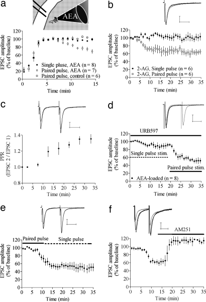
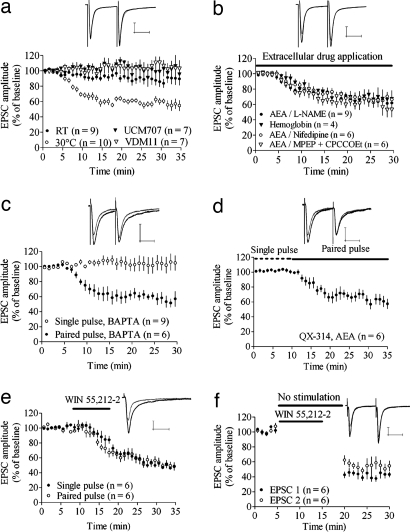
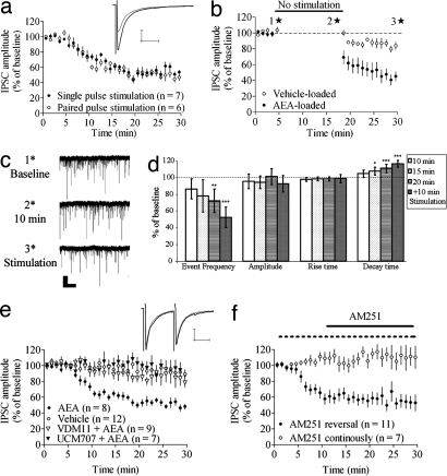
Endocannabinoids (eCBs) mediate short- and long-term depression of synaptic strength by retrograde transsynaptic signaling. Previous studies have suggested that an eCB mobilization or release step in the postsynaptic neuron is involved in this retrograde signaling. However, it is not known whether this release process occurs automatically upon eCB synthesis or whether it is regulated by other synaptic factors. To address this issue, we loaded postsynaptic striatal medium spiny neurons (MSNs) with the eCBs anandamide (AEA) or 2-arachidonoylglycerol and determined the conditions necessary for presynaptic inhibition. We found that presynaptic depression of glutamatergic excitatory postsynaptic currents (EPSCs) and GABAergic inhibitory postsynaptic currents (IPSCs) induced by postsynaptic eCB loading required a certain level of afferent activation that varied between the different synaptic types. Synaptic depression at excitatory synapses was temperature-dependent and blocked by the eCB membrane transport blockers, VDM11 and UCM707, but did not require activation of metabotropic glutamate receptors, l-calcium channels, nitric oxide, voltage-activated Na(+) channels, or intracellular calcium. Application of the CB(1)R antagonist, AM251, after depression was established, reversed the decrease in EPSC, but not in IPSC, amplitude. Direct activation of the CB(1) receptor by WIN 55,212-2 initiated synaptic depression that was independent of afferent stimulation. These findings indicate that retrograde eCB signaling requires a postsynaptic release step involving a transporter or carrier that is activated by afferent stimulation/synaptic activation.


Conflict of interest statement

The authors declare no conflict of interest.

Figures

Fig. 1.
Fig. 1.
eCB release depends on afferent activation. (a) Intracellular loading with AEA induced a depression in EPSC amplitude during paired-pulse, but not single-pulse, stimulation. The time for initial stabilization of EPSC amplitude after establishing the whole-cell recording was similar in AEA-loaded and control cells. Thus, the time courses for EPSC data in subsequent figures are shown starting from the time at which the response amplitude stabilized. (Inset) Schematic drawing of AEA loading into an MSN in the dorsolateral part of the striatum. Afferent activation was given through a bipolar electrode placed in the overlying white matter. (b) Postsynaptic loading of 2-AG induced a depression similar to that induced by AEA. (c) The depression in EPSC amplitude induced by AEA was concomitant with an increase in PPR. Normalized PPR values from nine cells are shown. (d) Treatment with 1 μM URB597, the FAAH blocker, slightly enhanced depression induced by AEA during single-pulse stimulation, but paired-pulse stimuli was required to induce a robust depression. (e) Paired-pulse stimulation was required for induction, but not maintenance, of AEA-induced depression (n = 6). (f) AEA-induced depression was completely reversed by 3 μM AM251, the CB1R antagonist (n = 5). Example trace shows baseline EPSC (black line) after t = 10–15 min (gray line) and after 10–15 min of AM251 treatment (dashed line). All other example traces show baseline EPSC (black lines) and after 20–25 min of single-pulse stimulation (gray lines). EPSC amplitude data are mean ± SEM. (Calibration bars, 25 msec and 100 pA.)
Fig. 2.
Fig. 2.
Molecular mechanisms of AEA loading-induced depression and differences relative to agonist-induced depression. (a) AEA-induced depression was blocked by postsynaptic loading of 10 μM VDM11 and 10 μM UCM707, the AMT blockers, and decreased at RT. Example traces show EPSCs at baseline and after 20–25 min of paired-pulse stimulation in MSNs loaded with AEA and UCM707. (b) Perfusion of 100 μM l-NAME, the NO synthetase inhibitor, or 500 mg/liter hemoglobin, the NO scavenger, did not prevent AEA-induced depression. EPSC amplitude also was depressed in AEA-loaded MSNs during treatment with 40 μM MPEP and 80 μM CPCCOEt, the group 1 mGluR antagonists, and during blockade of l-type calcium channels with 20 μM nifedipine. Example traces show EPSCs at baseline and after 20–25 min of paired-pulse stimulation during perfusion of l-NAME. (c) Chelation of intracellular calcium with 20 mM BAPTA did not prevent AEA-induced depression during paired-pulse stimulation and did not facilitate depression during single-pulse stimulation. (d) Blockade of voltage-gated sodium channels by 6 μM QX-314 did not affect AEA-induced depression. (e and f) Synaptic depression induced by activation of the CB1 receptor with extracellular WIN 55,212-2 is independent of afferent activation. Example traces show EPSC amplitude at the time point where paired-pulse stimulation started (t = 10 min) (black lines) and after 20–25 min of paired-pulse stimulation (gray lines). EPSC amplitude data are mean ± SEM. (Calibration bars, 25 msec and 100 pA.)
Fig. 3.
Fig. 3.
AEA-induced depression at GABAergic synapses. (a) AEA-induced depression at inhibitory synapses is independent of stimulation protocol. (b) IPSC amplitude also is depressed compared with baseline and vehicle when afferent activation is suspended but is further reduced after stimulation is resumed. (c) Representative traces showing sIPSCs at time points marked with stars in b. (Scale bar: 5 sec and 30 pA.) (d) Statistics for sIPSCs measured continuously in AEA-loaded MSNs, with afferent activation initiated 20 min after establishing the whole-cell recording configuration. Event frequency was significantly reduced after 20 min of postsynaptic AEA loading without afferent activation but decreased further subsequent to paired-pulse activation. (e) AEA-induced depression was prevented by postsynaptic loading of the AMT blockers VDM11 or UCM707. Example traces show IPSC at baseline and after 20–25 min of paired-pulse stimulation in MSNs loaded with AEA and UCM707. (f) Postsynaptic loading combined with afferent activation did not depress IPSCs during continuous AM251 perfusion but previously established depression was not reversed by a 20-min application of AM251. EPSC amplitude data are mean ± SEM. (Calibration bars, 25 msec and 100 pA.)

References

    1. Gerdeman G, Lovinger DM. J Neurophysiol. 2001;85:468–471. - PubMed
    1. Ronesi J, Gerdeman GL, Lovinger DM. J Neurosci. 2004;24:1673–1679. - PMC - PubMed
    1. Chevaleyre V, Takahashi KA, Castillo PE. Annu Rev Neurosci. 2006;29:37–76. - PubMed
    1. Alger BE, Pitler TA, Wagner JJ, Martin LA, Morishita W, Kirov SA, Lenz RA. J Physiol. 1996;496:197–209. - PMC - PubMed
    1. Pitler TA, Alger BE. Neuron. 1994;13:1447–1455. - PubMed

Publication types

MeSH terms

Substances

LinkOut - more resources