. 2007 Dec 26;104(52):20979-84.
doi: 10.1073/pnas.0707394104. Epub 2007 Dec 5.

Hematopoietic prostaglandin D2 synthase controls the onset and resolution of acute inflammation through PGD2 and 15-deoxyDelta12 14 PGJ2

Affiliations

Hematopoietic prostaglandin D2 synthase controls the onset and resolution of acute inflammation through PGD2 and 15-deoxyDelta12 14 PGJ2

Ravindra Rajakariar et al. Proc Natl Acad Sci U S A. .

Abstract

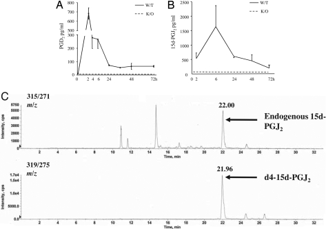
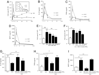
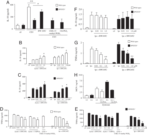
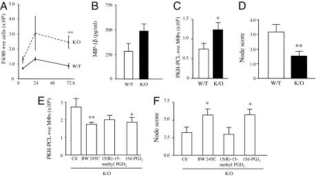
Hematopoietic prostaglandin D(2) synthase (hPGD(2)S) metabolizes cyclooxygenase (COX)-derived PGH(2) to PGD(2) and 15-deoxyDelta(12-14) PGJ(2) (15d-PGJ(2)). Unlike COX, the role of hPGD(2)S in host defense is ambiguous. PGD(2) can be either pro- or antiinflammatory depending on disease etiology, whereas the existence of 15d-PGJ(2) and its relevance to pathophysiology remain controversial. Herein, studies on hPGD(2)S KO mice reveal that 15d-PGJ(2) is synthesized in a self-resolving peritonitis, detected by using liquid chromatography-tandem MS. Together with PGD(2) working on its DP1 receptor, 15d-PGJ(2) controls the balance of pro- vs. antiinflammatory cytokines that regulate leukocyte influx and monocyte-derived macrophage efflux from the inflamed peritoneal cavity to draining lymph nodes leading to resolution. Specifically, inflammation in hPGD(2)S KOs is more severe during the onset phase arising from a substantial cytokine imbalance resulting in enhanced polymorphonuclear leukocyte and monocyte trafficking. Moreover, resolution is impaired, characterized by macrophage and surprisingly lymphocyte accumulation. Data from this work place hPGD(2)S at the center of controlling the onset and the resolution of acute inflammation where it acts as a crucial checkpoint controller of cytokine/chemokine synthesis as well as leukocyte influx and efflux. Here, we provide definitive proof that 15d-PGJ(2) is synthesized during mammalian inflammatory responses, and we highlight DP1 receptor activation as a potential antiinflammatory strategy.


Conflict of interest statement

The authors declare no conflict of interest.

Figures

Fig. 1.
Fig. 1.
hPGD2S synthesizes 15d-PGJ2 during zymosan-induced resolving peritonitis. (A and B) hPGD2S-derived PGD2 (A) and 15d-PGJ2 (B) were extracted from the cell-free inflammatory exudates and quantified by EIA and electrospray triple quadruple LC-MS/MS, respectively. Ex vivo degradation of PGD2 to 15d-PGJ2 during sample processing was controlled for by spiking inflammatory fluids in situ with deuterated PGD2. (C) LC-MS/MS spectra of detected 15d-PGJ2, which was found to be native and not deuterated. For eicosanoid analysis n = 6–8 animals were present in each group with the exception of 15d-PGJ2, which had 2–6 samples per group. Data are represented as the mean ± SEM.
Fig. 2.
Fig. 2.
PGD2 acting by the DP1 receptor controls the balance of pro- vs. antiinflammatory mediators and leukocyte trafficking during acute inflammation. (A) Intraperitoneal zymosan resulted in a self-limiting inflammatory response that peaked at 6–12 h. Inflammation in hPGD2S−/− mice (K/O) was 2-fold greater than in WT at 6 h and failed to resolve, with the early-onset phase characterized by predominantly GR1-positive PMNs with higher numbers of monocytes/macrophages compared with WT (see Fig. 4A). (Inset) Appropriate isotype control antibodies. (B–D) Cell-free exudate levels of IL-10 (B) were lower in hPGD2S−/− mice whereas TNFα (C) and MCP-1 (D) were higher. (E–I) The hyperinflammation in hPGD2S−/− mice was redressed by the DP1 receptor agonist BW245C (E) but not by the DP2 agonist 15(R)-15-methyl PGD2 (F), resulting in a corresponding (G–I) equilibration of inflammatory cytokines and chemokines to levels similar to controls. n = 6–8 animals per group; *, P ≤ 0.05; **, P ≤ 0.01, as determined by ANOVA followed by Bonferroni t test, with data expressed as mean ± SEM.
Fig. 3.
Fig. 3.
hPGD2S controls the inflammatory phenotype of peritoneal leukocytes. (A–C) IL-10 release from stimulated hPGD2S-deficient T cells (A) as well as B cells (B and C) was not only lower than that from wild types but was rescued by DP1 activation (BW245C). DP2 receptor activation with 15(R)-15-methyl PGD2 was without effect in these experiments. (D and E) Conversely, TNFα release from hPGD2S-deficient B cells was greater than that from wild types and was reversed with BW245C and 15d-PGJ2 but not 15(R)-15-methyl PGD2. (F–H) In macrophages, whereas hPGD2S had little effect on IL-10 (F), BW245C reduced TNFα (G) whereas 15d-PGJ2 also ameliorated levels of MCP-1 secretion (H). These data show the differential regulatory effects of hPGD2S-derived lipid mediators on inflammatory leukocyte phenotype. Cells were isolated from n = 3 animals, and all experiments done in triplicate on two separate occasions to confirm original findings. *, P ≤ 0.05; **, P ≤ 0.01, as determined by ANOVA, followed by Bonferroni t test, with data expressed as mean ± SEM.
Fig. 4.
Fig. 4.
hPGD2S deficiency compromises inflammatory resolution. (A and B) Although PMNs dominate the inflamed peritoneal cavity at the early-onset phase in hPGD2S−/− (K/O) mice (Fig. 2A, Inset) F4/80 positive macrophages were the predominant cell types during resolution (A) WT, wild type. Increased macrophage numbers in hPGD2S−/− mice could arise from enhanced MIP-1β (B) as well as MCP-1 (Fig. 2D) synthesis and/or a failure to exit the peritoneum to the parathymic lymph nodes (15, 16). To determine the latter, mice were injected i.p. with the macrophage label PKH-PCL26 at either 24 h or 48 h. (C) A significantly greater number of PKH26-PCL-positive macrophages (double labeling with FITC-labeled F4/80) were recorded in the cavity of hPGD2S−/− mice compared with WT at 72 h. (D) Correspondingly fewer labeled macrophages were found by histology in the parathymic lymph nodes of the KOs. (E and F) Adding BW245C (DP1 receptor agonist) or 15d-PGJ2 to the inflamed cavity of hPGD2S−/− mice reversed the accumulation of macrophages in the peritoneum (E) and resulted in increased numbers of labeled macrophages in parathymic lymph nodes (F). n = 10 animals per group; *, P ≤ 0.05; **, P ≤ 0.01, as determined by ANOVA, followed by Bonferroni t test, with data expressed as mean ± SEM.

Comment in

References

    1. Gilroy DW, Colville-Nash PR, Willis D, Chivers J, Paul-Clark MJ, Willoughby DA. Nat Med. 1999;5:698–701. - PubMed
    1. Gilroy DW, Newson J, Sawmynaden P, Willoughby DA, Croxtall JD. FASEB J. 2004;18:489–498. - PubMed
    1. Trivedi SG, Newson J, Rajakariar R, Jacques TS, Hannon R, Kanaoka Y, Eguchi N, Colville-Nash P, Gilroy DW. Proc Natl Acad Sci USA. 2006;103:5179–5184. - PMC - PubMed
    1. Urade Y, Hayaishi O. Vitam Horm. 2000;58:89–120. - PubMed
    1. Hammad H, de Heer HJ, Soullie T, Hoogsteden HC, Trottein F, Lambrecht BN. J Immunol. 2003;171:3936–3940. - PubMed

Publication types