Comparative Study
. 2007 Dec 11;104(50):19813-8.
doi: 10.1073/pnas.0708120104. Epub 2007 Dec 5.

Principles underlying energetic coupling along an allosteric communication trajectory of a voltage-activated K+ channel

Affiliations
Comparative Study

Principles underlying energetic coupling along an allosteric communication trajectory of a voltage-activated K+ channel

Evgeniya Sadovsky et al. Proc Natl Acad Sci U S A. .

Abstract

The information flow between distal elements of a protein may rely on allosteric communication trajectories lying along the protein's tertiary or quaternary structure. To unravel the underlying features of energy parsing along allosteric pathways in voltage-gated K(+) channels, high-order thermodynamic coupling analysis was performed. We report that such allosteric trajectories are functionally conserved and delineated by well defined boundaries. Moreover, allosteric trajectories assume a hierarchical organization whereby increasingly stronger layers of cooperative residue interactions act to ensure efficient and cooperative long-range coupling between distal channel regions. Such long-range communication is brought about by a coupling of local and global conformational changes, suggesting that the allosteric trajectory also corresponds to a pathway of physical deformation. Supported by theoretical analyses and analogy to studies analyzing the contribution of long-range residue coupling to protein stability, we propose that such experimentally derived trajectory features are a general property of allosterically regulated proteins.


Conflict of interest statement

The authors declare no conflict of interest.

Figures
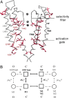
Fig. 1.
Fig. 1.
The allosteric communication network found in the pore domain of the Shaker Kv channel. (A) Gating-sensitive positions of the Shaker Kv channel pore (15), including the PVP activation gate residues (lower horizontal line), form a connected pattern when mapped onto the closed pore conformation of the KcsA K+ channel (for clarity, only two diagonal channel subunits are shown) (15). TM1, P, and TM2 denote the outer, pore, and inner helices, respectively. Side chains are shown only for gating sensitive residues (red) and for three gating-insensitive outer helix residues (gray; also indicated by asterisks) (15). The black arrow schematically represents a possible route for the allosteric trajectory of the Kv channel. The dashed gray and red arrows (left subunit) indicate the L403–T469 and E395–T469 residue pairs specifically discussed in the main text. (B) Double-mutant cycle analysis applied to voltage-dependent gating enables the measurement of state-dependent coupling. The middle cycle in bold and the cycle comprising the outer four corners represent the double-mutant cycles used to measure the coupling free energies between residues i and j in the closed (C) and open (O) states, respectively. M1, M2, and M1M2 denote the two single and double mutants, respectively. See SI Methods for further details.
Fig. 2.
Fig. 2.
Pairwise coupling free energies [Δ2G(i,j)] along the allosteric trajectory of the Kv channel are abolished upon perturbation of an adjacent trajectory residue. (A) Voltage–activation curves for four channel proteins comprising the double-mutant cycle measuring the coupling free energy between A391 and E395. (B) A thermodynamic cube is used to calculate the effect of a third residue, k, on the interaction between residues i and j (see SI Methods). (C and D) Voltage–activation curves for four channel proteins comprising the thermodynamic mutant cycle measuring the coupling free energy between A391 and E395 in the presence of the modified T469 (C) or A465 (D) trajectory-lining residues. Smooth curves correspond to a two-state Boltzmann function. The gray smooth curves correspond to the voltage–activation curves of the cycle presented in A. (E) Comparison of the magnitudes of coupling free energies [Δ2G(i,j)] between different trajectory-spanning residue pairs in the presence of a native (black bars) or mutated (gray bars) third trajectory-lining residue. The dashed gray lines delineate the 1 kcal/mol cutoff lines above which two residues are considered to be coupled (15).
Fig. 3.
Fig. 3.
Pairwise coupling energies along the allosteric trajectory are significantly reduced in the absence of an adjacent interaction. (A) A four-dimensional construct (double-mutant cycle of double-mutant cycles) is used to measure the effect of the interaction between T469 and A465 on the magnitude of coupling between the A391 and E395 residue pair (see SI Methods). (B) Voltage–activation curves for four channel proteins comprising a thermodynamic mutant cycle serve to calculate the coupling free energy between A391 and E395 in a T469A–A465V double-mutant background. (C) Comparison of the magnitude of coupling free energies [Δ2G(i, j)] between different trajectory-lining residue pairs in the presence (black bars) or absence (gray bars) of the indicated adjacent interactions.
Fig. 4.
Fig. 4.
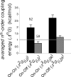
The boundaries of the allosteric trajectory are well defined. Presented here is a comparison of the averaged second- and third-order coupling free energies between trajectory-lining residue pairs and residue pairs comprising a trajectory-lining residue and an adjacent off-trajectory residue. The numbers indicated above the second-order coupling bars correspond to the average residue pair (side chain) distance for each group, calculated on the basis of the KcsA structure, after the appropriate residues had been mutated to those corresponding residues found in the Shaker channel. The values of Δ2G(i,j) and Δ3G(i,j)k used in this comparison are indicated in SI Table 2 and in table 2 of ref. .
Fig. 5.
Fig. 5.
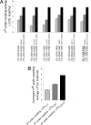
Allosteric communication trajectories exhibit increasingly stronger layers of cooperative interactions. (A) Comparison of the second-, third-, and fourth-order coupling free energies associated with the indicated residue pairs along the allosteric trajectory. For the third- and fourth-order coupling energies, the respective single residue or residue pair affecting the targeted (i, j) interaction is indicated in gray. (B) Comparison of the average high-order coupling free energies for the six trajectory interactions indicated in A. The high-order coupling free energies were calculated as described in SI Methods and are listed in SI Table 2.
Fig. 6.
Fig. 6.
High-order coupling free energy is related to cooperativity in channel gating. (A and B) The second- and third-order coupling free energies of the six trajectory residue pairs indicated in Fig. 5A were plotted as a function of the nonadditivity associated with either nH (A) or V1/2 (B) (see SI Methods). Negative values associated with nonadditivity in nH reflect the fact that nH effects are subadditive. (C) Comparison of the average high-order coupling free energies associated with the interactions presented in Fig. 5A, with the high-order nonadditivity associated with nH.

References

    1. Freire E. Proc Natl Acad Sci USA. 1999;96:10118–10122. - PMC - PubMed
    1. Pan H, Lee JC, Hilser VJ. Proc Natl Acad Sci USA. 2000;97:12020–12025. - PMC - PubMed
    1. Liu T, Whitten ST, Hilser VJ. Proteins. 2006;62:728–738. - PubMed
    1. Lockless SW, Ranganathan R. Science. 1999;286:295–299. - PubMed
    1. Suel GM, Lockless SW, Wall MA, Ranganathan R. Nat Struct Biol. 2003;10:59–69. - PubMed

Publication types

MeSH terms

Substances