

Comparative Study
doi: 10.1073/pnas.0709915104.
Epub 2007 Dec 5.
Localizing frustration in native proteins and protein assemblies
Affiliations
- PMID: 18077414
- PMCID: PMC2148382
- DOI: 10.1073/pnas.0709915104
Item in Clipboard
Comparative Study
Localizing frustration in native proteins and protein assemblies
Proc Natl Acad Sci U S A.
.
Abstract
We propose a method of quantifying the degree of frustration manifested by spatially local interactions in protein biomolecules. This method of localization smoothly generalizes the global criterion for an energy landscape to be funneled to the native state, which is in keeping with the principle of minimal frustration. A survey of the structural database shows that natural proteins are multiply connected by a web of local interactions that are individually minimally frustrated. In contrast, highly frustrated interactions are found clustered on the surface, often near binding sites. These binding sites become less frustrated upon complex formation.
Conflict of interest statement
The authors declare no conflict of interest.
Figures
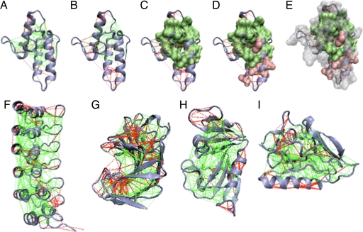
Examples of the localized frustration and minimally frustrated networks in protein structures. The protein backbone, direct interresidue interactions, and water-mediated interactions are displayed as blue ribbons, solid lines, and dashed lines, respectively. Minimally frustrated interactions and highly frustrated ones are shown in green and red, respectively; neutral contacts are not drawn. The surfaces indicate the single-residue-level frustration index, using a corresponding coloring scheme. (A) Im7 protein [Protein Data Bank (PDB) ID code 7CEI] mutational frustration index. (B) Configurational frustration index. (C–E) Single-residue frustration index surfaces overlaid. (F–I) Configurational frustration index of IκBα (PDB ID code 1NFI) (F), Streptomyces Endoglucanase (PDB ID code 1OA4) (G), dihydrofolate reductase (PDB ID code 1RX2) (H), and Endostatin (PDB ID code 1KOE) (I).
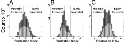
Histograms showing the distribution of the mutational frustration index (light gray) and the configurational frustration index (dark gray) in the three different contact classes considered. These frustration indices were computed for every native contact present in a database of 314 monomeric protein domains. (A) Short-range contacts. (B) Long-range contacts. (C) Water-mediated contacts. The vertical lines indicate the energy landscape theory-based cutoff used to define minimally frustrated interactions as well as the cutoff for neutral or highly frustrated interactions.
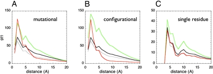
The pair distribution functions between the centers of mass of the contacts was calculated for a database of 314 monomeric proteins (black). The distributions for minimally frustrated (green), neutral (gray), or highly frustrated contacts (red) are shown both for each class using the mutational frustration index (A), the configurational frustration index (B), or the single-residue-level frustration index (C). For the single-residue-level frustration index, the distance between Cα atoms was used.
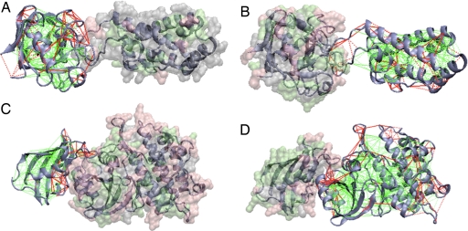
Examples of localized frustration patterns in protein assemblies. The interactions in one monomeric partner are colored according to the contact configurational frustration index, whereas the other partner's surface is colored according to the single-residue-level frustration index, as described for Fig. 1. The frustration indices are shown as calculated for the unbound monomers. Complementary views of the same complexes are shown and correspond to the database members cyclophilin bound to the N-terminal domain of HIV-1 capsid (PDB ID code 1AK4) (A and B) and cytoplasmic domain of the type 1 TGF-β receptor in complex with fkbp12 (PDB ID code 1B6C) (C and D).
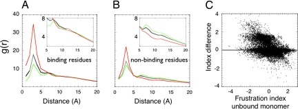
Frustration distribution in protein assemblies. (A and B) The pair distribution functions between the center of mass of contacts classified by their frustration index (minimally frustrated in green, neutral in gray, or highly frustrated in red) to the Cα of either binding (A) or the surface residues not involved in binding (B) are shown. (C) The change of the configurational frustration index upon binding for the contacts close to the binding site in all of the complexes analyzed.
References
-
- Bryngelson JD, Wolynes PG. J Phys Chem. 1989;93:6902–6915.
-
- Clementi C, Maritan A, Banavar JR. Phys Rev Lett. 1998;81:3287–3290.
Publication types
MeSH terms
Substances
Grants and funding
LinkOut - more resources
Full Text Sources
