Comparative Study
. 2007 Dec 11;104(50):19831-6.
doi: 10.1073/pnas.0709978104. Epub 2007 Dec 5.

How processing of aspartylphosphate is coupled to lumenal gating of the ion pathway in the calcium pump

Affiliations
Comparative Study

How processing of aspartylphosphate is coupled to lumenal gating of the ion pathway in the calcium pump

Chikashi Toyoshima et al. Proc Natl Acad Sci U S A. .

Abstract

Ca(2+)-ATPase of skeletal muscle sarcoplasmic reticulum is the best-studied member of the P-type or E1/E2 type ion transporting ATPases. It has been crystallized in seven different states that cover nearly the entire reaction cycle. Here we describe the structure of this ATPase complexed with phosphate analogs BeF(3)(-) and AlF(4)(-) in the absence of Ca(2+), which correspond to the E2P ground state and E2 approximately P transition state, respectively. The luminal gate is open with BeF(3)(-) and closed with AlF(4)(-). These and the E1 approximately P.ADP analog crystal structures show that a two-step rotation of the cytoplasmic A-domain opens and closes the luminal gate through the movements of the M1-M4 transmembrane helices. There are several conformational switches coupled to the rotation, and the one in the cytoplasmic part of M2 has critical importance. In the second step of rotation, positioning of one water molecule couples the hydrolysis of aspartylphosphate to closing of the gate.


Conflict of interest statement

The authors declare no conflict of interest.

Figures

Fig. 1.
Fig. 1.
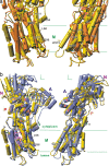
Structure of the BeF3 complex of Ca2+-ATPase in the absence of Ca2+ and a comparison of the cytoplasmic domains with other phosphorylated intermediate analogs. (a) Ribbon model of E2· BeF3(−TG), with several marker residues in ball and stick. The image was prepared with Molscript (33). (b) Details around the phosphorylation site in E2·BeF3(TG). (c) Positions of the A-domain relative to the P-domain in E1·AlF4·ADP (orange), E2·BeF3(−TG) (light green), E2·BeF3(TG) (cyan, transparent), and E2·AlF4(TG) (blue gray). The P-domain is shown only for E2·BeF3(TG). The purple net in b represents an ∣Fo∣ − ∣Fc∣ electron density map contoured at 6σ at 2.4-Å resolution before introducing BeF3 and Mg2+ into the atomic model. Rotation axes for E1·AlF4 ADP → E2·BeF3(−TG) (I) and E2·BeF3(−TG) → E2·AlF4(TG) (II) are specified in a and c. In b, pink broken lines show likely hydrogen bonds and the white arrow a van der Waals contact.
Fig. 2.
Fig. 2.
Superimposition of the models for phosphorylated intermediate analogs fitted with the M7–M10 helices. Helices are represented with cylinders with marker residues in stick. The colors used are yellow, E2·BeF3(−TG); orange, E1·AlF4·ADP; and blue gray, E2·AlF4(TG). Small arrows show movements in the transition from E1P → E2P (a) and E2P → E2∼P (b). The TG binding site is indicated with an inverted triangle (b).
Fig. 3.
Fig. 3.
Cross-sections of the transmembrane region of Ca2+-ATPase. van der Waals surfaces of E1·AlF4·ADP (a) and E2·BeF3(−TG) (b). Red colors represent acidic residues. Dotted circles in b indicate the positions of Ca2+ observed in E1·AlF4·ADP (a). The images shown were prepared with PyMol (34).
Fig. 4.
Fig. 4.
Atomic models fitted with the A-domain, illustrating the M2 switch. Cα traces of the A-domain and the M1–M2 helices (Asp-59–Thr-242) are shown in stereo with side chains of several marker residues. The colors used are orange, E1·AlF4·ADP; yellow, E2·BeF3(−TG); cyan, E2·BeF3(TG); blue gray, E2·AlF4(TG); and lime, E2(TG+BHQ) (also marked). Arrows represent the distances between Asn-111 and Ala-115 in E1·AlF4·ADP and E2·AlF4(TG).
Fig. 5.
Fig. 5.
Details of the phosphorylation site in the phosphorylated intermediate analogs. (a) E2·BeF3(TG), E2P ground state analog. (b) E2·AlF4(TG), E2∼P transition state analog. (c) E2·MgF42(TG) (PDB ID code 1WPG), E2·Pi product state analog. Broken lines in pink show likely hydrogen bonds, and those in light green show Mg2+ coordination. Small red spheres represent water molecules. The conserved sequence motifs are shown.
Fig. 6.
Fig. 6.
Surface representation of the atomic models for the phosphorylation site in the E2P and E2∼P analogs. The van der Waals surface is superimposed on the atomic model of E2·BeF3(TG) (a) or E2·AlF4(TG) (b) with explicit hydrogens. Green spheres represent the atoms in the A-domain; yellow spheres represent those in the P-domain. Phosphate analogs (dimensions taken from phosphate), Mg2+ (blue violet spheres), and water (red and cyan spheres) are shown in atom colors. The white arrow in b indicates the attacking water molecule. Thr-353 is located on the front side and not seen here.
Fig. 7.
Fig. 7.
A cartoon illustrating two-step rotation in the processing of aspartylphosphate and gating of the ion pathway. Small arrows indicate the movements of the TM helices. The M1–M4 (green) and A1–A3 (yellow) helices are numbered. P-D351 refers to phosphorylated Asp-351.

References

    1. MacLennan DH, Brandl CJ, Korczak B, Green NM. Nature. 1985;316:696–700. - PubMed
    1. de Meis L, Vianna AL. Annu Rev Biochem. 1979;48:275–292. - PubMed
    1. Albers RW. Annu Rev Biochem. 1967;36:727–756. - PubMed
    1. Post RL, Hegyvary C, Kume S. J Biol Chem. 1972;247:6530–6540. - PubMed
    1. Toyoshima C, Nakasako M, Nomura H, Ogawa H. Nature. 2000;405:647–655. - PubMed

Publication types

MeSH terms

LinkOut - more resources