. 2007 Dec 11;104(50):20114-9.
doi: 10.1073/pnas.0707626104. Epub 2007 Dec 6.

Skeletal muscle resists stretch by rapid binding of the second motor domain of myosin to actin

Affiliations

Skeletal muscle resists stretch by rapid binding of the second motor domain of myosin to actin

Elisabetta Brunello et al. Proc Natl Acad Sci U S A. .

Abstract

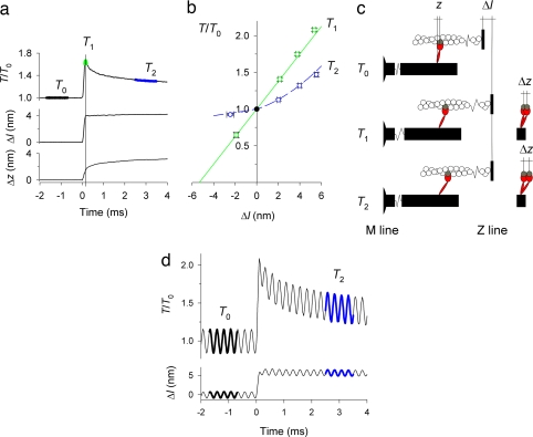
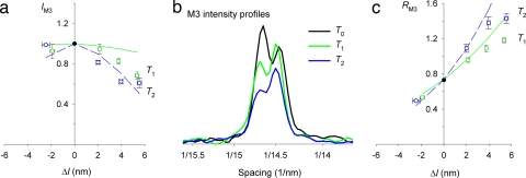
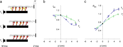
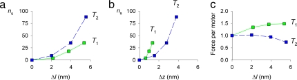
A shortening muscle is a machine that converts metabolic energy into mechanical work, but, when a muscle is stretched, it acts as a brake, generating a high resistive force at low metabolic cost. The braking action of muscle can be activated with remarkable speed, as when the leg extensor muscles rapidly decelerate the body at the end of a jump. Here we used time-resolved x-ray and mechanical measurements on isolated muscle cells to elucidate the molecular basis of muscle braking and its rapid control. We show that a stretch of only 5 nm between each overlapping set of myosin and actin filaments in a muscle sarcomere is sufficient to double the number of myosin motors attached to actin within a few milliseconds. Each myosin molecule has two motor domains, only one of which is attached to actin during shortening or activation at constant length. A stretch strains the attached motor domain, and we propose that combined steric and mechanical coupling between the two domains promotes attachment of the second motor domain. This mechanism allows skeletal muscle to resist external stretch without increasing the force per motor and provides an answer to the longstanding question of the functional role of the dimeric structure of muscle myosin.


Conflict of interest statement

The authors declare no conflict of interest.

Figures

Fig. 1.
Fig. 1.
Mechanical response to a step stretch imposed during isometric contraction. (a) Time course of force (T) relative to isometric force (T0), length change of the half-sarcomere (Δl), and axial motion of myosin motors (Δz); time of x-ray exposures at T0, T1, and T2 indicated on the force trace. (b) T1 (green) and T2 (blue) plotted against Δl (mean ± SEM, n = 19 fibers); data for shortening steps, here and elsewhere, are from ref. . The green straight line was obtained by regression of the T1 data for ≈2-nm shortening and stretch steps and the isometric point (T0, black circle); the blue dashed line was drawn through the T2 points by eye. T0 was 285 ± 90 kPa (mean ± SD). (c) (Left) Myosin motor (red) with its catalytic domain attached to a monomer (gray) in the actin filament (white) and light-chain domain (LCD) attached to the myosin filament backbone (black). Axial motion after a stretch is accompanied by tilting of the LCD and quantified by the axial separation z between the two ends of the LCD. (Right) Motor conformations at T1 and T2 superimposed on that at T0. (d) Force response (upper trace) to a 5.7-nm stretch with superimposed 4-kHz oscillations (lower trace). Force and length oscillations used for the analysis are indicated by thickening of the traces. Black indicates T0; blue indicates T2.
Fig. 2.
Fig. 2.
Structural response to step stretch. (a) Intensity of the M3 x-ray reflection (IM3). Symbols are the same as in Fig. 1b. Lines are from the structural model in ref. . Green indicates T1; blue indicates T2. (b) Intensity profiles of M3 reflection along the meridional axis. Colors are the same as in Fig. 1. (c) Changes in the relative intensity of the two major peaks of the M3 reflection (RM3). Symbols and lines are the same as in a.
Fig. 3.
Fig. 3.
Structural model including stretch-induced attachment of myosin motors. (a) Structural model of motors and filaments modified from Fig. 1c. Red indicates motors attached to actin monomers (dark gray) at T0, yellow indicates detached partner motors, and pink indicates partner motors attached to the next actin monomer on the M-ward side (light gray). (b and c) IM3 (b) and RM3 (c) data from Fig. 2 a and c, respectively. Lines are from the model with the stretch-induced attachment described in the text. Symbol and color codes are as in Fig. 2a.
Fig. 4.
Fig. 4.
Number (ns) of additional myosin motors attached to actin after a stretch and the force per motor. (a) ns plotted against stretch size (Δl). (b) ns plotted against axial motion of motors (Δz). (c) Average force per myosin motor plotted against Δl. Green indicates T1; blue indicates T2.

References

    1. Katz B. J Physiol (London) 1939;96:45. - PMC - PubMed
    1. Infante AA, Klaupiks D, Davies RE. Science. 1964;144:1577–1578. - PubMed
    1. Curtin NA, Davies RE. Cold Spring Harbor Symp Quant Biol. 1973;37:619–626.
    1. Linari M, Woledge RC, Curtin NA. J Physiol (London) 2003;548:461–474. - PMC - PubMed
    1. Linari M, Lucii L, Reconditi M, Casoni ME, Amenitsch H, Bernstorff S, Piazzesi G, Lombardi V. J Physiol (London) 2000;526:589–596. - PMC - PubMed

Publication types