. 2008 Feb;19(2):735-44.
doi: 10.1091/mbc.e07-09-0968. Epub 2007 Dec 12.

Arx1 is a nuclear export receptor for the 60S ribosomal subunit in yeast

Affiliations

Arx1 is a nuclear export receptor for the 60S ribosomal subunit in yeast

Nai-Jung Hung et al. Mol Biol Cell. 2008 Feb.

Abstract

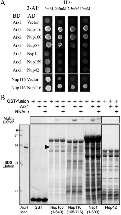
We previously showed that nuclear export of the large (60S) ribosomal subunit relies on Nmd3 in a Crm1-dependent manner. Recently the general mRNA export factor, the Mtr2/Mex67 heterodimer, was shown to act as an export receptor in parallel with Crm1. These observations raise the possibility that nuclear export of the 60S subunit in Saccharomyces cerevisiae requires multiple export receptors. Here, we show that the previously characterized 60S subunit biogenesis factor, Arx1, also acts as an export receptor for the 60S subunit. We found that deletion of ARX1 was synthetic lethal with nmd3 and mtr2 mutants and was synthetic sick with several nucleoporin mutants. Deletion of ARX1 led to accumulation of pre-60S particles in the nucleus that were enriched for Nmd3, Crm1, Mex67, and Mtr2, suggesting that in the absence of Arx1, 60S export is impaired even though the subunit is loaded with export receptors. Finally, Arx1 interacted with several nucleoporins in yeast two-hybrid as well as in vitro assays. These results show that Arx1 can directly bridge the interaction between the pre-60S particle and the NPC and thus is a third export receptor for the 60S subunit in yeast.


Figures

Figure 1.
Figure 1.
nmd3ΔC14 is trapped in the nucleus and impairs 60S subunit export. (A) Wild-type (WT) and nmd3ΔC14 strains were cultured in YPD to midlog phase and subjected to indirect immunofluorescence for visualization of endogenous Nmd3 localization. The effect of nmd3ΔC14 on nuclear export of the 60S subunit was monitored by Rpl25-eGFP. The nmd3l505a allele has an effect similar to that of nmd3ΔC14 on blocking nuclear export of the 60S subunit, as monitored by Rpl25-eGFP. (B) Cartoon of the primary structure of Nmd3. CC: Cys-x2-Cys repeats that comprise zinc-binding motifs. Dark gray indicate regions to which mutations that disrupt 60S binding map (Hedges et al., 2006). Amino acid sequence of the C-terminus of Nmd3 with the residues deleted in the nmd3ΔC14 mutant shown in light gray. An alignment of NESs of Nmd3 from various organisms is shown. The canonical leucine-rich NES sequence is shown with two examples from PKI and HIV-1 Rev. (Φ, hydrophobic residues (L,M,I,V); X, any amino acid). Gray bars highlight conserved residues. Sc, S. cerevisiae; Dm, Drosophila melanogaster; and Hs, Homo sapiens. Numbers indicate amino acid positions.
Figure 2.
Figure 2.
Elimination of Arx1 impairs nuclear export of Nmd3 and 60S subunits in the nup120ΔC16 strain. (A) Functional analysis of regulatable Arx1-myc constructs. arx1Δ nup120ΔC16 cells expressing either wild-type Arx1-myc (pAJ1026), Gal-UBI(M)-Arx1-myc (pAJ1481) or Gal-UBI(R)-Arx1-myc (pAJ1484) plasmid as the sole copy of Arx1 were tested for growth on galactose- or glucose-containing dropout medium. (B) Protein expression of regulatable Arx1-myc expression constructs. The arx1Δ nup120ΔC16 cells harboring either Arx1-myc, Gal-UBI(M)-Arx1-myc (pAJ1481) or Gal-UBI(R)-Arx1-myc (pAJ1484) plasmid were cultured in galactose-containing media to midlog phase and transferred to glucose-containing media for the indicated times before they were collected. Extracts were subjected to SDS-PAGE and Western blotting using anti-myc antibody. (C) The arx1Δ nup120ΔC16 stain carrying the unstable Gal-UBI(R)-Arx1-myc construct was transformed with either Rpl25-eGFP (pAJ908) or Nmd3-GFP (pAJ755) plasmid. The strains were then cultured in galactose containing media to midlog phase and transferred to glucose containing media for the indicated times before they were subjected to microscopy. Nuclear DNA was stained with DAPI.
Figure 3.
Figure 3.
Nmd3-bound 60S particles accumulate in the nucleus in an arx1Δ mutant. (A) Cultures of wild-type and arx1Δ cells carrying pAJ582 (Nmd3-GFP) were grown to midlog phase in selective media and the localization of Nmd3 was monitored by fluorescence microscopy. (B) Lysates were prepared from wild-type and arx1Δ cells and fractioned on 7–47% sucrose gradients by ultracentrifugation as described in Materials and Methods. Fractions were collected, and the absorbance at 254 nm was monitored continuously. Proteins were precipitated with trichloroacetic acid, separated by SDS-PAGE, transferred to nitrocellulose membrane, and immunoblotted for Nmd3 or Rpl8p using specific antibodies.
Figure 4.
Figure 4.
Deletion of Arx1 leads to nuclear accumulation of an Nmd3-Crm1–60S complex. (A) Wild-type and arx1Δ cells transformed with pAJ538 (Nmd3-13myc) or pAJ1594 (Nmd3-supraNES-13myc) and pAJ739 (Crm1T539C-HA) were cultured in selective media and collected at midlog phase. Immunoprecipitations were carried out using anti-myc antibodies and subjected to Western blotting using anti-myc and anti-HA antibodies to monitor Nmd3 and Crm1 levels. N/A, negative control. (B) Lysates from wild-type and arx1Δ cells carrying pAJ856 (Crm1-HA) were prepared in the presence of cycloheximide and fractioned on 7–47% sucrose gradients by ultracentrifugation. Fractions were collected, and the absorbance at 254 nm was monitored continuously. Proteins were precipitated with trichloroacetic acid, separated by SDS-PAGE, transferred to nitrocellulose membrane, and immunoblotted for HA or Rpl8p using specific antibodies.
Figure 5.
Figure 5.
Mex67 and Mtr2 are enriched on pre-60S particles when export is blocked. Cell extracts were prepared from wild-type (BY4741) and arx1Δ (AJY1901) expressing Nmd3-myc (pAJ538) and Nmd3(AAA)-myc (pAJ752). The myc-tagged Nmd3 proteins were immunoprecipitated¤ and proteins that copurifying proteins were detected by Western blotting using antibodies against Mex67, Mtr2, Nmd3-myc, and Rpl8, as a marker for 60S subunits.
Figure 6.
Figure 6.
ARX1 shows genetic interactions with other 60S export factors. (A) An arx1Δ mutant was mated to mtr2-33 containing MTR2 on a URA3 vector. After sporulation and dissecting, serial dilutions of spore clones of the indicated genotypes were spotted onto 5FOA media to select against the wild-type MTR2 vector. Plates were incubated at 30°C for 4 d. Additional results from high copy expression and synthetic effects of double mutants are summarized in Table 4. (B) NMD3, MEX67, MTR2¤ and CRM1 were expressed from high copy vectors in wild-type (BY4741) and arx1Δ mutant cells (AJY1901). Vector indicates an empty vector control. Serial dilutions were plated onto selective media and incubated for 3 d at 30°C.
Figure 7.
Figure 7.
Arx1 interacts with nucleoporins. (A) A panel of plasmids that express Arx1 or the Nup116 FG domain, as a control, fused to the Gal4-binding domain (BD) and various Nup FG domains fused to the Gal4 activation domain (AD) were transformed into yeast strain BJ69-4A and spotted onto selective media (Leu Ura His dropout for Arx1-Nup interactions or Leu Trp His dropout for Nup-Nup interactions) to test for potential interactions. Plates were incubated at 30°C for 4 d. Positive interactions should drive expression of the HIS3 reporter. The indicated concentrations of 3-AT were added to increase the stringency of the assay. (B) In vitro interaction of Arx1 with GST-Nup fusions. GST, GST-Nup100(aa 1–640), GST-Nup116(aa 165–716) and GST-Nsp1(aa 1–603)-coated beads (2 μg GST fusion protein in 10 μl of beads) were incubated with purified Arx1 (0.8 μg) in the absence or presence of RNase A (25 μg). After 1 h at 4°C, beads were concentrated by centrifugation, washed twice, and bound proteins eluted first with 0.3 M MgCl2, followed by elution with SDS. Bound proteins were resolved by SDS-PAGE and visualized with Coomassie blue. The bands present below the full-length GST-Nup fusion proteins in the lanes without Arx1 are degradation products of the fusion proteins.

Similar articles

Cited by

References

    1. Allen N. P., Huang L., Burlingame A., Rexach M. Proteomic analysis of nucleoporin interacting proteins. J. Biol. Chem. 2001;276:29268–29274. - PubMed
    1. Allen N. P., Patel S. S., Huang L., Chalkley R. J., Burlingame A., Lutzmann M., Hurt E. C., Rexach M. Deciphering networks of protein interactions at the nuclear pore complex. Mol. Cell Proteom. 2002;1:930–946. - PubMed
    1. Bassler J., Grandi P., Gadal O., Lessmann T., Petfalski E., Tollervey D., Lechner J., Hurt E. Identification of a 60S preribosomal particle that is closely linked to nuclear export. Mol. Cell. 2001;8:517–529. - PubMed
    1. Belaya K., Tollervey D., Kos M. FLIPing heterokaryons to analyze nucleo-cytoplasmic shuttling of yeast proteins. RNA. 2006;12:921–930. - PMC - PubMed
    1. Bischoff F. R., Klebe C., Kretschmer J., Wittinghofer A., Ponstingl H. RanGAP1 induces GTPase activity of nuclear Ras-related Ran. Proc. Natl. Acad. Sci. USA. 1994;91:2587–2591. - PMC - PubMed

Publication types

MeSH terms

Substances