. 2008 Feb;19(2):711-21.
doi: 10.1091/mbc.e07-07-0713. Epub 2007 Dec 12.

Maintenance of hormone-sensitive phosphoinositide pools in the plasma membrane requires phosphatidylinositol 4-kinase IIIalpha

Affiliations

Maintenance of hormone-sensitive phosphoinositide pools in the plasma membrane requires phosphatidylinositol 4-kinase IIIalpha

Andras Balla et al. Mol Biol Cell. 2008 Feb.

Abstract

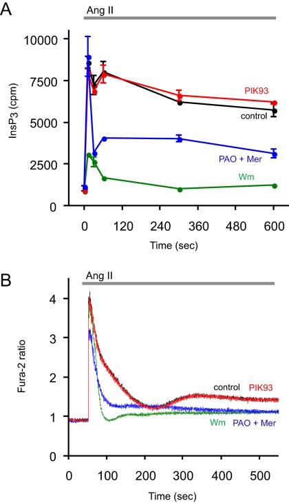
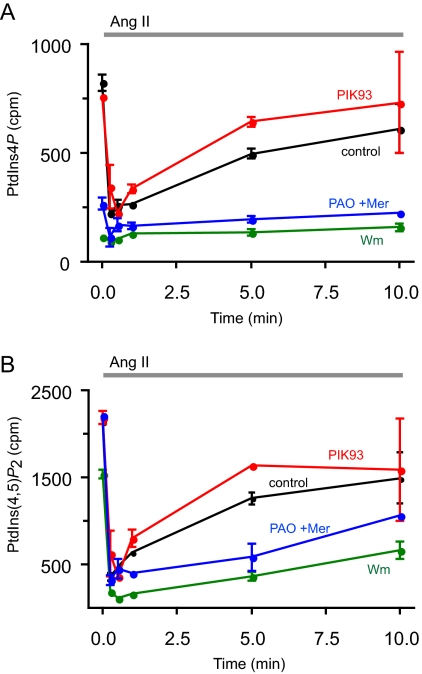
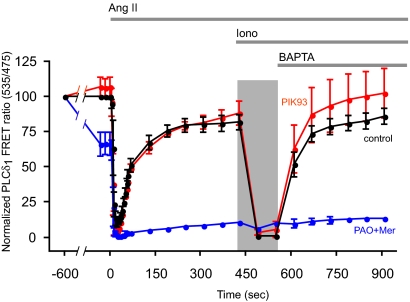
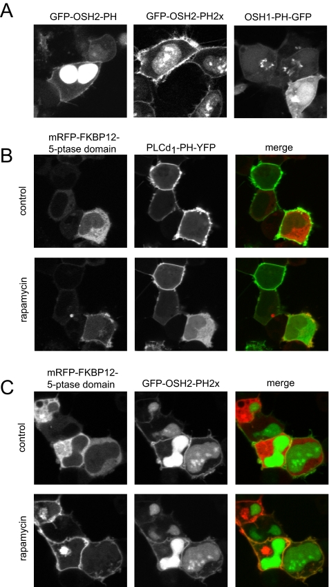
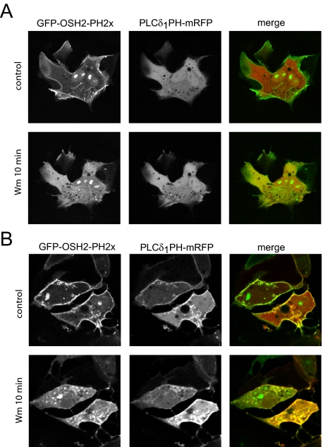
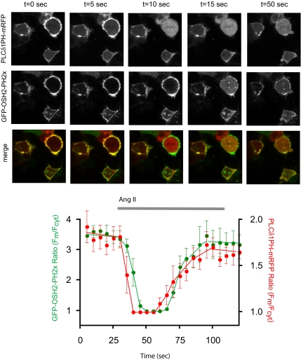
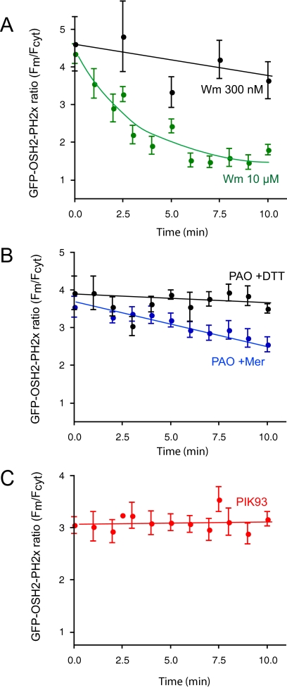
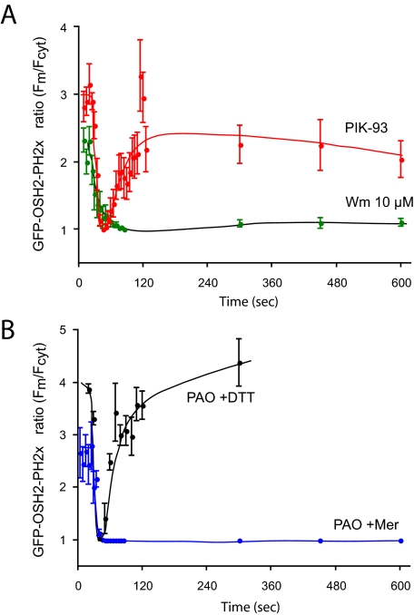
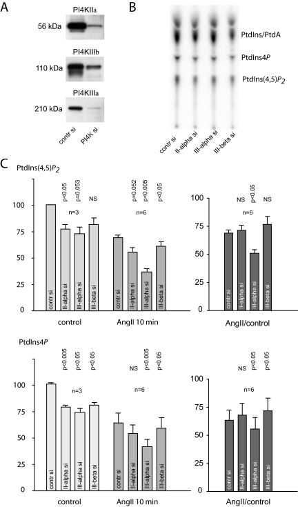
Type III phosphatidylinositol (PtdIns) 4-kinases (PI4Ks) have been previously shown to support plasma membrane phosphoinositide synthesis during phospholipase C activation and Ca(2+) signaling. Here, we use biochemical and imaging tools to monitor phosphoinositide changes in the plasma membrane in combination with pharmacological and genetic approaches to determine which of the type III PI4Ks (alpha or beta) is responsible for supplying phosphoinositides during agonist-induced Ca(2+) signaling. Using inhibitors that discriminate between the alpha- and beta-isoforms of type III PI4Ks, PI4KIIIalpha was found indispensable for the production of phosphatidylinositol 4-phosphate (PtdIns4P), phosphatidylinositol 4,5-bisphosphate [PtdIns(4,5)P(2)], and Ca(2+) signaling in angiotensin II (AngII)-stimulated cells. Down-regulation of either the type II or type III PI4K enzymes by small interfering RNA (siRNA) had small but significant effects on basal PtdIns4P and PtdIns(4,5)P(2) levels in (32)P-labeled cells, but only PI4KIIIalpha down-regulation caused a slight impairment of PtdIns4P and PtdIns(4,5)P(2) resynthesis in AngII-stimulated cells. None of the PI4K siRNA treatments had a measurable effect on AngII-induced Ca(2+) signaling. These results indicate that a small fraction of the cellular PI4K activity is sufficient to maintain plasma membrane phosphoinositide pools, and they demonstrate the value of the pharmacological approach in revealing the pivotal role of PI4KIIIalpha enzyme in maintaining plasma membrane phosphoinositides.


Figures

Figure 1.
Figure 1.
Effects of PI4K inhibitors on the kinetics of InsP3 and [Ca2+]i changes in HEK-293-AT1 cells stimulated with AngII. (A) HEK-293-AT1 cells were labeled with myo-[3H]inositol for 24 h in inositol-free medium as described under Materials and Methods. After washing, AngII (10−7 M) was added to the cells for the indicated times, and reactions were terminated by PCA. Labeled inositol phosphates were extracted from the soluble fraction and separated by high-performance liquid chromatography connected to a scintillation flow detector as described previously (Nakanishi et al., 1995). Data are shown are means ± range of duplicate determinations. The concentrations of the inhibitors added 10 min before AngII were 250 nM PIK93, 10 μM PAO with 1 mM β-mercaptoethanol (Mer), and 10 μM Wm. Two additional experiments were performed with the 10-min time points with similar results. (B) HEK-293-AT1 cells were loaded with Fura-2/AM, and their fluorescence monitored in a fluorescent spectrophotometer as described under Materials and Methods. AngII (10−7 M) was added at the indicated time. Pretreatment with the inhibitors for 10 min was as described in A. This result is a representative of three similar observations.
Figure 2.
Figure 2.
Effects of PI4K inhibitors on the kinetics of phosphoinositide changes in HEK-293-AT1 cells stimulated with AngII. HEK-293-AT1 cells were labeled with [32P]phosphate for 3 h in a phosphate-free medium as described under Materials and Methods. AngII (10−7 M) was added to the cells at the indicated times, and reactions terminated by the addition of PCA. Lipids were extracted from the cell pellets, separated by TLC, and analyzed both by a PhosphorImager and scintillation counting of the spots cut out from the plates. Data shown are means ± range of duplicate determinations. The concentrations of the inhibitors added 10 min before AngII were 250 nM PIK93, 10 μM PAO with 1 mM Mer, and 10 μM Wm. This full time course experiment was repeated from myo-[3H]inositol-labeled cells with similar results, supporting the same conclusion.
Figure 3.
Figure 3.
Effects of PI4K inhibitors on the kinetics of redistribution of PLCδ1PH domain in HEK-293-AT1 cells stimulated with AngII. HEK-293 cells stably transfected with the AT1a angiotensin receptors were cotransfected with the PLCd1PH-CFP and -YFP constructs for 24 h. The FRET signal from individual cells was then monitored in a wide-field fluorescent microscope equipped with an emission beam splitter using 430-nm excitation and 535- and 475-nm emissions as described under Materials and Methods. To minimize light exposure, after the first minute of stimulation, data were collected in longer intervals. AngII (10−7 M) was added at the indicated time followed by 10 μM inonomycin (Iono) and 5 mM BAPTA (or EGTA). The dark bar represents the exposure of the cells to the high (extracellular) Ca2+ concentration after ionomycin treatment. The concentrations of the inhibitors added 10 min before AngII were 250 nM PIK93 and 10 μM PAO with 1 mM Mer. FRET was expressed as fluorescent emission ratio values (535/475 nm), and it was normalized so that the ratio value recorded before the addition of inhibitors was taken as 100% and the lowest values (after AngII or ionomycin) were taken as 0%. Means ± SEM (or range) from five, two, and three cells are shown for control, PAO + Mer, and PIK93, respectively. Similar changes were observed in two additional experiments.
Figure 4.
Figure 4.
Localization of OSH1- and OSH2-PH domain-GFP fusion proteins in HEK-293-AT1 cells. (A) The PH domains of the yeast oxysterol-binding protein homologues OSH2 and OSH1 were fused to GFP as described under Materials and Methods. These constructs were transfected into HEK-293-AT1 cells and examined 24 h after transfection with live cell confocal microscopy. Note that the OSH2-PH domain binds to the plasma membrane and strongly accumulates in the nucleus but does not bind to the Golgi. A tandem PH domain of the OSH2 protein shows smaller signal in the nucleus and a strong plasma membrane binding. The OSH1-PH domain binds both the plasma membrane and the Golgi. (B) Elimination of the plasma membrane localization of PLCδ1PH-GFP [monitoring PtdIns(4,5)P2] by plasma membrane recruitment of a phosphoinositide 5-phosphatase. HEK-293-AT1 cells were transfected with a truncated type-IV phosphoinositide 5-phosphatase fused to mRFP and FKBP12, a plasma membrane-targeted FRB-CFP construct and the PLCδ1PH-YFP reporter described in Varnai et al. (2006). Addition of rapamycin for 3 min recruits the otherwise cytoplasmic 5-ptase construct to the plasma membrane (left) with a concomitant elimination of PtdIns(4,5)P2 and loss of PLCδ1PH-YFP localization (middle). (C) The same manipulations do not eliminate the plasma membrane localization of the OSH2-PH2x-GFP, suggesting that this construct is not kept at the membrane by PtdIns(4,5)P2.
Figure 5.
Figure 5.
Localization of OSH2-PH2x-GFP to the plasma membrane is wortmannin sensitive. (A) COS-7 cells were transfected with OSH2-PH2x-GFP together with PLCδ1PH-mRFP and the wild-type type IV phosphoinositide 5-phosphatase for 24 h. Cells were selected so that the PLCδ1PH-mRFP showed no localization, indicating the lack of PtdIns(4,5)P2 as a result of phosphatase expression. These cells still showed plasma membrane localization of OSH2-PH2x-GFP, indicating that the construct is kept in the membrane not by PtdIns(4,5)P2. Addition of 10 μM Wm to such cells caused a rapid translocation of the OSH2-PH2x-GFP domain construct from the membrane to the cytosol. (B) Release of the OSH2-PH2x-GFP construct from the membrane after Wm treatment is significantly slower in control cells where PtdIns(4,5)P2 is present in the membrane.
Figure 6.
Figure 6.
Angiotensin II-induced changes in membrane phosphoinositides simultaneously monitored by OSH2-PH2x-GFP and PLCδ1PH-mRFP. HEK-293 cells stably transfected with the AT1a angiotensin receptors were cotransfected with the PLCδ1PH-mRFP and OSH2-PH2x-GFP constructs for 24 h. Live cells were examined in a confocal microscope at 35°C during stimulation with AngII (10−7 M), and pictures were taken every 5 s. Note the faster response observed with the PLCδ1PH-mRFP. Quantification of membrane/cytosolic fluorescence intensities as function of time calculated from line-intensity histograms from five to seven cells obtained in two separate experiments (means ± SEM).
Figure 7.
Figure 7.
Effects of PI4K inhibitors on the membrane localization of the OSH2-PH2x-GFP. HEK-293 cells stably transfected with the AT1a angiotensin receptors were cotransfected with the OSH2-PH2x-GFP constructs for 24 h. Live cells were examined in a confocal microscope at 35°C. Calculation of membrane/cytosolic fluorescence intensities as function of time was performed from line-intensity histograms from images taken at every minute during a 10-min incubation with the inhibitors. The concentrations of the inhibitors were 250 nM PIK93, 10 μM PAO with 1 mM Mer, 10 μM PAO with 1 mM DTT, and 10 μM or 300 nM Wm. Means ± SEM from three to 25 cells from two to four separate experiments are shown.
Figure 8.
Figure 8.
Effects of AngII on the membrane localization of the OSH2-PH2x-GFP after treatment with PI4K inhibitors. HEK-293 cells stably transfected with the AT1a angiotensin receptors were cotransfected with the OSH2-PH2x-GFP constructs for 24 h. Live cells were examined in a confocal microscope at 35°C. Calculation of membrane/cytosolic fluorescence intensities as function of time was performed from line-intensity histograms from images taken at the indicated times after AngII addition. The concentrations of the inhibitors applied for 10 min before AngII were 250 nM PIK93, 10 μM PAO with 1 mM Mer, and 10 μM Wm. Means ± SEM from three to 25 cells from two to four separate experiments are shown. The initial ratio values of Wm-treated cells are higher in these experiments than those shown in Figure 7 at the end of Wm treatment, because cells with still measurable OSH2-PH2x-GFP localization after Wm treatment were selected for this analysis.
Figure 9.
Figure 9.
Effects of knockdown of the individual PI 4-kinases on [32P]phosphate labeling of phosphoinositides and their response to AngII stimulation in HEK-293-AT1 cells. HEK-293 cells stably transfected with the AT1a receptors were treated with siRNA for 3 d as described under Materials and Methods. The extent of knockdown was determined by Western blot analysis (A). On the fourth day, cells were labeled with [32P]phosphate for 3 h in phosphate-free medium. Cells were then either treated with AngII (10−7 M) or saline and incubated for an additional 10 min. Reactions were terminated by PCA, and lipids were extracted from the cell pellets, separated by TLC, and analyzed by a PhosphorImager (B). Due to cell loss in the PI4K-depleted cells, the individual PtdIns4P and PtdIns(4,5)P2 spots were normalized to the PtdA/PtdIns values measured in unstimulated samples of the respective groups. These ratios were then compared between the PI4K down-regulated or control siRNA-treated cells in each of the three experiments performed in duplicates (C). In each experiments, the effect of 250 nM PIK93 was also determined in the AngII group. Because PIK93-treated cells showed no difference compared with those treated with AngII only, these values were pooled in the statistical analysis. Means ± SEM are shown, and the p values obtained in one-sample t tests compared with control siRNA-treated cells are shown above the bars.

References

    1. Audhya A., Emr S. D. Stt4 PI 4-kinase localizes to the plasma membrane and functions in the Pkc1-mediated MAP kinase cascade. Dev. Cell. 2002;2:593–605. - PubMed
    1. Audhya A., Foti M., Emr S. D. Distinct roles for the yeast phosphatidylinositol 4-kinases, stt4p and pik1p, in secretion, cell growth, and organelle membrane dynamics. Mol. Biol. Cell. 2000;11:2673–2689. - PMC - PubMed
    1. Balla A., Balla T. Phosphatidylinositol 4-kinases; old enzymes with emerging functions. Trends Cell Biol. 2006;16:351–361. - PubMed
    1. Balla A., Tuymetova G., Barshishat M., Geiszt M., Balla T. Characterization of type II phosphatidylinositol 4-kinase isoforms reveals association of the enzymes with endosomal vesicular compartments. J. Biol. Chem. 2002;277:20041–22050. - PubMed
    1. Balla A., Tuymetova G., Tsiomenko A., Varnai P., Balla T. A plasma membrane pool of phosphatidylinositol 4-phosphate is generated by phosphatidylinositol 4-kinase type-III alpha: studies with the PH domains of the oxysterol binding protein and FAPP1. Mol. Biol. Cell. 2005;16:1282–1295. - PMC - PubMed

Publication types

MeSH terms