. 2008 Feb;99(2):976-88.
doi: 10.1152/jn.00930.2007. Epub 2007 Dec 12.

Circadian- and light-dependent regulation of resting membrane potential and spontaneous action potential firing of Drosophila circadian pacemaker neurons

Affiliations

Circadian- and light-dependent regulation of resting membrane potential and spontaneous action potential firing of Drosophila circadian pacemaker neurons

Vasu Sheeba et al. J Neurophysiol. 2008 Feb.

Abstract

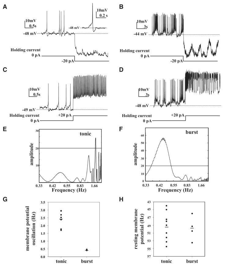
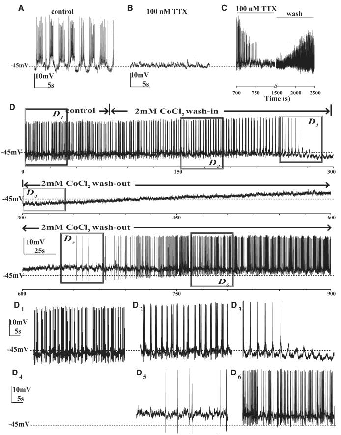
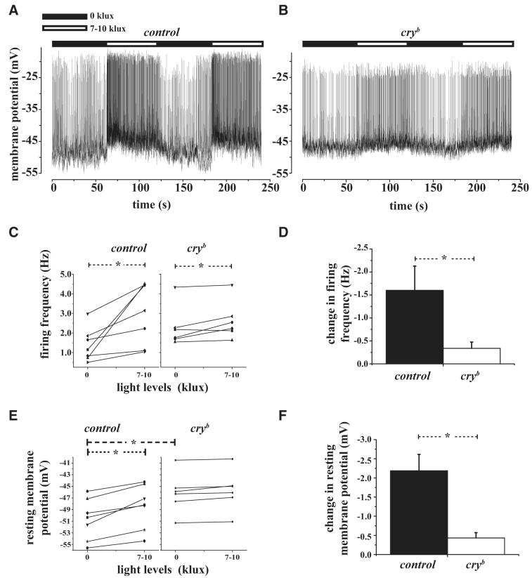
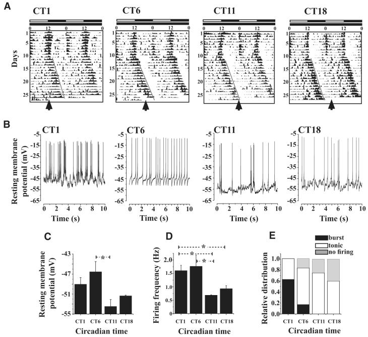
The ventral lateral neurons (LNvs) of adult Drosophila brain express oscillating clock proteins and regulate circadian behavior. Whole cell current-clamp recordings of large LNvs in freshly dissected Drosophila whole brain preparations reveal two spontaneous activity patterns that correlate with two underlying patterns of oscillating membrane potential: tonic and burst firing of sodium-dependent action potentials. Resting membrane potential and spontaneous action potential firing are rapidly and reversibly regulated by acute changes in light intensity. The LNv electrophysiological light response is attenuated, but not abolished, in cry(b) mutant flies hypomorphic for the cell-autonomous light-sensing protein CRYPTOCHROME. The electrical activity of the large LNv is circadian regulated, as shown by significantly higher resting membrane potential and frequency of spontaneous action potential firing rate and burst firing pattern during circadian subjective day relative to subjective night. The circadian regulation of membrane potential, spontaneous action potential firing frequency, and pattern of Drosophila large LNvs closely resemble mammalian circadian neuron electrical characteristics, suggesting a general evolutionary conservation of both physiological and molecular oscillator mechanisms in pacemaker neurons.


Figures

Fig. 1
Fig. 1
Spontaneous tonic and burst firing action potentials of large ventral lateral (LNv) pacemaker neurons. A: representative trace of spontaneous action potentials measured by whole cell recording from a large LNv of freshly dissected brain from an adult fly showing tonic firing pattern. Spontaneous changes in membrane potential were measured in current-clamp configuration with 0-pA holding current. Hyperpolarizing current injection (-20 pA) step from 0-pA holding current blocks spontaneous firing. At 0 holding current spontaneous action potentials (APs) show gradual depolarization approaching spike threshold, then a depolarizing spike followed by repolarization and afterhyperpolarization (inset: detail of single tonic AP). Tonic firing large LNv resting membrane potential is -49 ± 1 mV, n = 9. B: representative traces of large LNv spontaneous exhibiting burst pattern of AP firing measured in current-clamp configuration with 0-pA holding current followed by hyperpolarization (-20 pA) that blocks AP firing but not membrane potential oscillations. C: depolarizing current injection (+20 pA) step from 0 pA holding current increases tonic firing AP firing rate. D: depolarizing (+20 pA) current injection increases firing rate during burst firing. Mean resting membrane potential during burst firing is -50 ± 2 mV (mean ± SE, n = 4). E and F: representative periodograms obtained by Lomb—Scargle time-series analysis during tonic firing showing a single statistically significant period with frequency of 1.76 Hz (frequency range for tonic firing is 1.7 to 3.0 Hz, n = 9) and burst firing that shows a significant spectral peak around 0.45 Hz (frequency range for burst firing is 0.4 to 0.47 Hz, n = 4). During burst firing large LNvs exhibit low-frequency (0.49 ± 0.07 Hz, n = 4), high-amplitude (7 ± 1 mV, n = 4) slow oscillations in membrane potential that underlie the burst firing. G: scatterplot of frequency of oscillation in membrane potential as measured by Lomb—Scargle analysis during tonic and burst firing modes showing a higher range during tonic firing and no frequency overlap between tonic and burst firing modes. H: mean resting membrane potential during burst firing is -50 ± 2 mV (mean ± SE, n = 4), which does not differ significantly from tonic firing mode, as shown by scatterplot comparison.
Fig. 2
Fig. 2
Voltage-gated sodium and calcium channel blockers abolish the spontaneous AP firing of large LNvs. A: firing of a representative large LNv measured in whole cell current clamp at 0-pA holding membrane current in control perfusion solution. B: recording from the same cell in the presence of 100 nM tetrodotoxin (TTX). The application of 100 nM TTX completely abolishes large LNv spontaneous APs (n = 6). C: example trace showing recovery of spontaneous firing after washout of TTX (2/6 cells). D: representative long-duration whole cell current-clamp recording of one large LNv at 0-pA holding current in control solution during washin and washout of 2 mM CoCl2 shows state modulation of a large LNv showing burst firing becoming tonic and then silent with 2 mM CoCl2 washin followed by washout recovery to tonic and then burst firing pattern (x-axis shows time in seconds, n = 6). D1–6: detail of traces sampled from the above trace for 20 s. D1: burst firing before CoCl2 washin, (D2) initial broadening of slow-wave oscillation and crests of firing in response to CoCl2 washin, (D3) during transition from tonic to silent, (D4) silent phase, (D5) transition from silent to tonic showing recovery of large LNv spontaneous firing after CoCl2 washout, and (D6) transition from tonic to burst firing showing complete washout.
Fig. 3
Fig. 3
Spontaneous AP firing of large LNv increase rapidly in response to light in a CRYPTOCHROME (CRY)-dependent manner. A: representative large LNv whole cell current-clamp recording from control large LNv showing depolarized resting membrane potential and increased spontaneous AP firing rate within seconds in response to light increase from 0 to 7–10 klux (light levels indicated by the horizontal bars above each trace: black bar, lights-off; white bar, lights-on) where light levels were at 0 lux (off) during the 1st and 3rd min and increased to 7–10 klux (on) during the 2nd and 4th min of the recording. Control large LNv cells show depolarized resting membrane potential and increased spontaneous AP firing rate in response to light and hyperpolarized resting membrane potential and decreased spontaneous AP firing in response to the cessation of light. B: representative trace from cryb mutant flies recorded under similar condition as control. C: mean spontaneous AP firing frequency increases significantly from 0 to 7–10 klux light levels as determined by paired t-test for large LNv control (P = 0.01) and cryb mutant flies (P = 0.02). D: the light response is not detectable in cryb mutant large LNv cells because increase in spontaneous AP firing frequency is significantly higher for controls compared with cryb mutant flies (t-test, P = 0.03). E: large LNv resting membrane potential becomes significantly more depolarized in response to lights-on for controls (paired t-test P = 0.001) but not in cryb mutant flies. The baseline resting membrane potential in cryb mutant large LNv at 0 klux is significantly greater than controls. F: the relative change in resting membrane potential is significantly greater in controls compared with cryb mutant flies (t-test, P = 0.002).
Fig. 4
Fig. 4
Electrophysiological properties of large LNv cells exhibit circadian oscillation. A: representative locomotor activity/rest records of individual flies maintained under 12 h/12 h light/dark (LD) cycle for 10 days following which they were transferred to constant darkness (DD). During LD, lights remained on from 0 to 12 h as indicated by the white horizontal bar and remained off for the next 12 h as indicated by the black horizontal bar. The activity/rest recordis double plotted to enable better visualization of the circadian rhythm. Locomotor activity synchronizes to the imposed LD cycle as indicated by the increased levels of activity around lights-on and -off and relative quiescence during night and midday. After release into DD, the activity/rest rhythms “free-runs” with a period slightly longer than 24 h, reflecting the endogenous period of circadian clock of each individual fly. By convention the offset of activity is referred to as circadian time CT12 and was used as a phase marker in our experiments. Flies were sampled at 4 distinct phases, CT1, 6, 11, and 18, based on the predicted offset of activity after 15 days of DD as indicated by an arrow. B: representative current-clamp traces showing that large LNv spontaneous AP firing frequency and resting membrane potential values are higher during subjective day compared with night phases (sample sizes for CT1, 6, 11, and 18 were 8, 6, 4, and 5 brains, respectively). All recordings were made under identical low-light conditions of 0.1 klux. C: mean resting membrane potential is highest at CT6 and lowest at CT11 (one-way ANOVA, P = 0.0246; * indicates significant differences, Tukey’s HSD, CT6 vs. CT11, P = 0.02). D: large LNv firing frequency is significantly highest at CT6 and least at CT11 (one-way ANOVA, P = 0.045; * indicates significant differences, Fisher’s least-significant difference test; P < 0.05; CT6 vs. CT11, P = 0.025; CT6 vs. CT18, P = 0.04; CT1 vs. CT11, P = 0.04). E: burst firing pattern predominates at CT1. Patterns of firing were classified as tonic or bursting based on the frequencies obtained using Lomb—Scargle periodogram analysis and verified by visual examination of the traces. No burst firing is observed at CT18. The greatest number of instances of cells that do not fire spontaneous APs occur at CT18 (although nonfiring large LNv mean resting potentials recorded at this time point are within the normal range relative to tonic firing mode).

References

    1. Cloues RK, Sather WA. Afterhyperpolarization regulates firing rate in neurons of the suprachiasmatic nucleus. J Neurosci. 2003;23:1593–1604. - PMC - PubMed
    1. Collins B, Mazzoni EO, Stanewsky R, Blau J. Drosophila CRYPTOCHROME is a circadian transcriptional repressor. Curr Biol. 2006;16:441–449. - PubMed
    1. Colwell CS. Circadian modulation of calcium levels in cells in the suprachiasmatic nucleus. Eur J Neurosci. 2000;12:571–576. - PMC - PubMed
    1. de Jeu M, Hermes M, Pennartz C. Circadian modulation of membrane properties in slices of rat suprachiasmatic nucleus. Neuroreport. 1998;9:3725–3729. - PubMed
    1. de Jeu M, Pennartz C. Circadian modulation of GABA function in the rat suprachiasmatic nucleus: excitatory effects during the night phase. J Neurophysiol. 2002;87:834–844. - PubMed

Publication types

MeSH terms