. 2007 Dec 12;27(50):13624-34.
doi: 10.1523/JNEUROSCI.2858-07.2007.

The neonatal ventromedial hypothalamus transcriptome reveals novel markers with spatially distinct patterning

Affiliations

The neonatal ventromedial hypothalamus transcriptome reveals novel markers with spatially distinct patterning

Deborah M Kurrasch et al. J Neurosci. .

Abstract

The ventromedial hypothalamus (VMH) is a distinct morphological nucleus involved in feeding, fear, thermoregulation, and sexual activity. It is essentially unknown how VMH circuits underlying these innate responses develop, in part because the VMH remains poorly defined at a cellular and molecular level. Specifically, there is a paucity of cell-type-specific genetic markers with which to identify neuronal subgroups and manipulate development and signaling in vivo. Using gene profiling, we now identify approximately 200 genes highly enriched in neonatal (postnatal day 0) mouse VMH tissue. Analyses of these VMH markers by real or virtual (Allen Brain Atlas; http://www.brain-map.org) experiments revealed distinct regional patterning within the newly formed VMH. Top neonatal markers include transcriptional regulators such as Vgll2, SF-1, Sox14, Satb2, Fezf1, Dax1, Nkx2-2, and COUP-TFII, but interestingly, the highest expressed VMH transcript, the transcriptional coregulator Vgll2, is completely absent in older animals. Collective results from zebrafish knockdown experiments and from cellular studies suggest that a subset of these VMH markers will be important for hypothalamic development and will be downstream of SF-1, a critical factor for normal VMH differentiation. We show that at least one VMH marker, the AT-rich binding protein Satb2, was responsive to the loss of leptin signaling (Lep(ob/ob)) at postnatal day 0 but not in the adult, suggesting that some VMH transcriptional programs might be influenced by fetal or early postnatal environments. Our study describing this comprehensive "VMH transcriptome" provides a novel molecular toolkit to probe further the genetic basis of innate neuroendocrine behavioral responses.


Figures
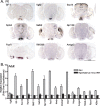
Figure 1.
Figure 1.
Expression patterns and levels of top VMH markers. In situ hybridization of top VMH markers in P0 embryos and qPCR in adult. A, Expression of novel VMH markers in P0 mouse pups is shown for coronal brain sections (20 μm thick). The pattern for the current definitive VMH marker, SF-1, is shown in the top left panel. Anatomical landmarks include the third ventricle (3V) and the amygdala (A). Scale bar, 2 mm. B, Relative transcript levels are shown for several VMH markers in adult male mice (>8 weeks). Relative levels of each transcript were assayed in the VMH (gray bars) and in the hypothalamus minus the VMH (black bars). Fold induction is relative to the hypothalamus minus the VMH and taken to be 1 (horizontal dashed line), with one of four control samples randomly set to equal 1. All other samples, including additional control and experimental samples, are normalized to this one value (or 1 tissue). The variation among each group of tissues is shown by error bars.
Figure 2.
Figure 2.
Expression of Vgll2 is present in the embryo and postnatal VMH but is absent in the early adult. In situ hybridization of Vgll2 and SF-1 in E12, P0, P7, and P21 brains. Vgll2 (top) and SF-1 (bottom) expression is shown for coronal brain sections (20 μm). The VMH is marked with a dashed oval, and the third ventricle (3V) is labeled for orientation.
Figure 3.
Figure 3.
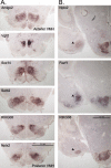
Expression patterns of new markers reveal spatially distinct expression patterns in the VMH, with some expressed in the medial amygdala. A, Expression patterns are shown for six different VMH markers in serial coronal brain sections obtained from P0 male mice (20 μm) beginning with the most anterior section (Amigo2, top) and extending posterior (Nptx2, bottom). Scale bar, 0.5 mm. B, Expression of Nptx2, Fezf1, and RIKG08 in the medial amygdala (arrowhead) is shown relative to expression in the VMH (for other genes found to be expressed in the VMH and in the amygdala, see supplemental Fig. S2, available at www.jneurosci.org as supplemental material). Scale bar, 1.0 mm.
Figure 4.
Figure 4.
Knockdown of VMH markers disrupts medial hypothalamic patterning in developing zebrafish. A, Diagram depicting the spatial patterning of known hypothalamic markers and VMH-specific makers in the 48 hpf zebrafish brain. Whole-mount in situ hybridization expression profiles are shown for orthologs of mouse neonatal VMH-enriched markers. Other anatomical landmarks include the developing eye and yolk sac. Images represent the most ventral side of the embryo with the dorsal side down. B, Whole-mount expression patterns for wild-type and morpholino-injected 48 hpf zebrafish are shown for nr5a1a (top 5 panels), nr5a2 (middle 5 panels), or hypocretin (bottom 5 panels). Sequences for all antisense morpholinos, including nmbr-MO, sox14-MO, a2bp1-MO, and fezf1-MO, are provided in supplemental Table S3 (available at www.jneurosci.org as supplemental material). Disruption of the nr5a1a and nr5a2 expression are noted by the lack of a discernable third ventricle boundary (arrowhead). Images are displayed from no phenotype (nmbr-MO), to weakly disrupted (fezf1-MO), and to severely disrupted (sox14-MO). MA, Mandibular arch. For each condition, two distinct morpholinos directed against the initiator methionine or splice junction/5′ UTR were used independently and together. Data shown are representative of two separate experiments using at least 80 embryos.
Figure 5.
Figure 5.
Expression of VMH-enriched transcripts requires SF-1. A, Expression patterns for Fezf1, Nkx2-2, and A2bp1 are shown in sf-1−/− P0 male mice. For comparison, SF-1 is shown (top). Expression of these markers is maintained in other brain regions (arrowheads). B, Relative transcript levels of VMH-enriched genes are shown for sf-1−/− mice (whole hypothalamus); expression in wild-type (WT) mice for each transcript is set at 100% (data not shown). C, Relative luciferase activity (RLU) is shown after transfection of wild-type or mutant promoter plasmids (100 ng; Materials and Methods) and increasing amounts of pCMV–SF-1 (0, 100, and 200 ng).
Figure 6.
Figure 6.
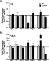
Reduction of Satb2 in the neonatal but not adult VMH. Relative levels of VMH transcripts as well as ARC transcripts, NPY, and POMC, as indicated on the x-axis are shown from isolated total hypothalami obtained from neonatal (P0; A) and adult (B) mice. Levels for wild-type (gray bars) and for Lepob/ob (black bars) are shown in both panels. Fold induction is relative to the hypothalamus minus the VMH with the wild-type (WT) control sample assigned a value of 1 (horizontal dashed line).

References

    1. Altman J, Bayer SA. The development of the rat hypothalamus. Adv Anat Embryol Cell Biol. 1986;100:1–178. - PubMed
    1. Barker DJ, Winter PD, Osmond C, Margetts B, Simmonds SJ. Weight in infancy and death from ischaemic heart disease. Lancet. 1989;2:577–580. - PubMed
    1. Bates SH, Stearns WH, Dundon TA, Schubert M, Tso AW, Wang Y, Banks AS, Lavery HJ, Haq AK, Maratos-Flier E, Neel BG, Schwartz MW, Myers MG., Jr STAT3 signalling is required for leptin regulation of energy balance but not reproduction. Nature. 2003;421:856–859. - PubMed
    1. Berghuis P, Rajnicek AM, Morozov YM, Ross RA, Mulder J, Urban GM, Monory K, Marsicano G, Matteoli M, Canty A, Irving AJ, Katona I, Yanagawa Y, Rakic P, Lutz B, Mackie K, Harkany T. Hardwiring the brain: endocannabinoids shape neuronal connectivity. Science. 2007;316:1212–1216. - PubMed
    1. Bjartmar L, Huberman AD, Ullian EM, Renteria RC, Liu X, Xu W, Prezioso J, Susman MW, Stellwagen D, Stokes CC, Cho R, Worley P, Malenka RC, Ball S, Peachey NS, Copenhagen D, Chapman B, Nakamoto M, Barres BA, Perin MS. Neuronal pentraxins mediate synaptic refinement in the developing visual system. J Neurosci. 2006;26:6269–6281. - PMC - PubMed

Publication types

MeSH terms