. 2007 Dec 12;27(50):13680-90.
doi: 10.1523/JNEUROSCI.2203-07.2007.

TRPM8 mechanism of cold allodynia after chronic nerve injury

Affiliations

TRPM8 mechanism of cold allodynia after chronic nerve injury

Hong Xing et al. J Neurosci. .

Abstract

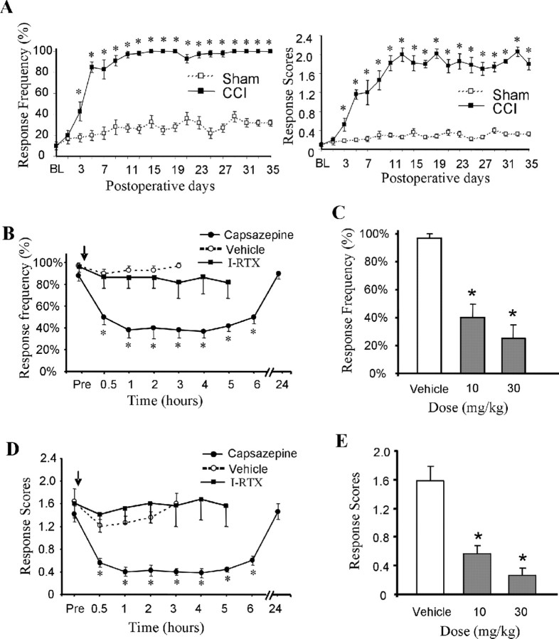
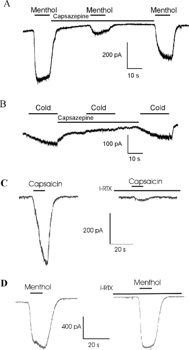
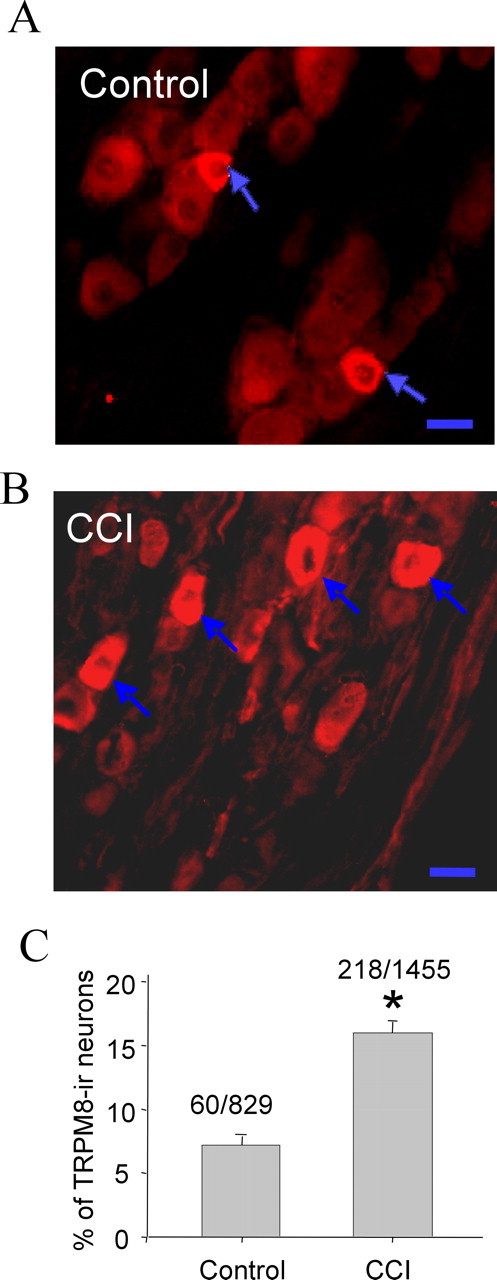
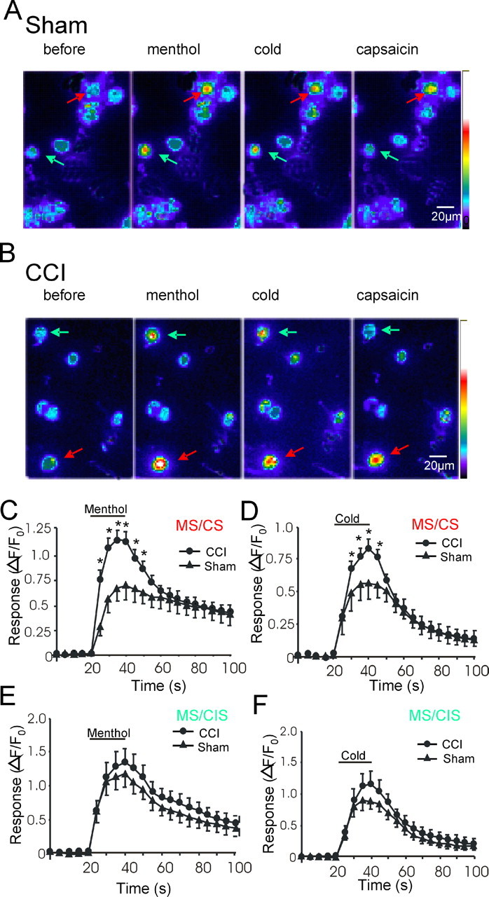
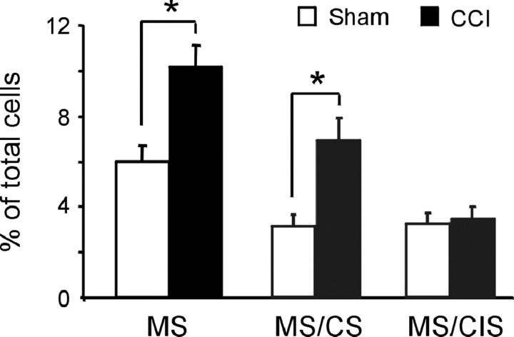
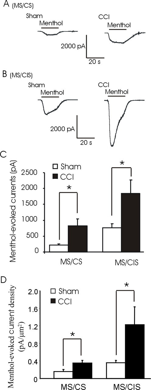
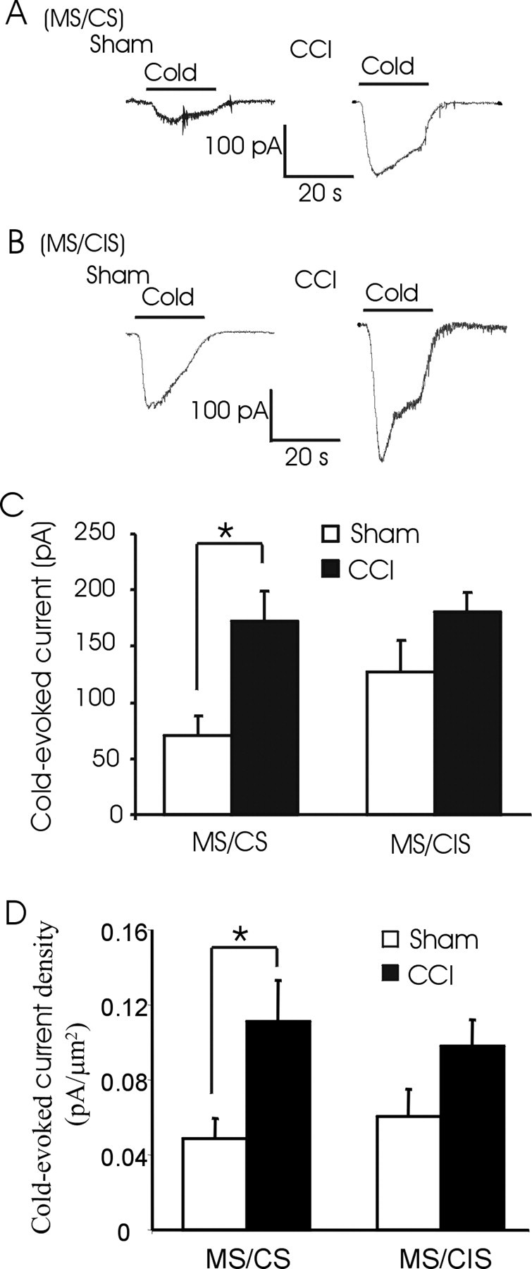
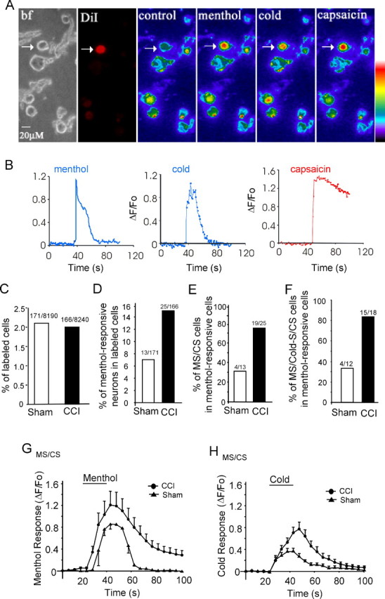
The cold- and menthol-sensitive receptor TRPM8 (transient receptor potential melastatin 8) has been suggested to play a role in cold allodynia, an intractable pain seen clinically. We studied how TRPM8 is involved in cold allodynia using rats with chronic constrictive nerve injury (CCI), a neuropathic pain model manifesting cold allodynia in hindlimbs. We found that cold allodynic response in the CCI animals was significantly attenuated by capsazepine, a blocker for both TRPM8 and TRPV1 (transient receptor potential vanilloid 1) receptors, but not by the selective TRPV1 antagonist I-RTX (5-iodoresiniferatoxin). In L5 dorsal root ganglion (DRG) sections of the CCI rats, immunostaining showed an increase in the percentage of TRPM8-immunoreactive neurons when compared with the sham group. Using the Ca2+-imaging technique and neurons acutely dissociated from the L5 DRGs, we found that CCI resulted in a significant increase in the percentage of menthol- and cold-sensitive neurons and also a substantial enhancement in the responsiveness of these neurons to both menthol and innocuous cold. These changes occurred in capsaicin-sensitive neurons, a subpopulation of nociceptive-like neurons. Using patch-clamp recordings, we found that membrane currents evoked by both menthol and innocuous cold were significantly enhanced in the CCI group compared with the sham group. By retrograde labeling afferent neurons that target hindlimb skin, we showed that the skin neurons expressed TRPM8 receptors, that the percentage of menthol-sensitive/cold-sensitive/capsaicin-sensitive neurons increased, and that the menthol- and cold-evoked responses were significantly enhanced in capsaicin-sensitive neurons after CCI. Together, the gain of TRPM8-mediated cold sensitivity on nociceptive afferent neurons provides a mechanism of cold allodynia.


Figures

Figure 1.
Figure 1.
Attenuation of cold allodynic responses by capsazepine in the CCI model. A, Time course of cold allodynic responses to the acetone test in ipsilateral hindpaws of the CCI rats (filled squares; n = 10) and the sham rats (open squares; n = 8). The responses are expressed either as response frequency (left) or response scores (right). BL, Baseline. B, Time course of cold allodynic response frequency in the CCI rats before and after an intraperitoneal single-dose injection of 10 mg/kg capsazepine (filled circles; n = 12), 0.75 mg/kg I-RTX (filled squares; n = 4), and vehicle (open circles; n = 6). The arrow indicates the injections of the testing compounds or vehicle. C, Summary of cold allodynic response frequency after the administration of vehicle (open bar; n = 6), 10 mg/kg capsazepine (first filled bar; n = 12), and 30 mg/kg capsazepine (second filled bar; n = 6). D, Time course of cold allodynic response scores in the CCI rats before and after an intraperitoneal single-dose injection of 10 mg/kg capsazepine (filled circles; n = 12), 0.75 mg/kg I-RTX (filled squares; n = 4), and vehicle (open circles; n = 6). The arrow indicates the injections of the testing compounds or vehicle. E, Summary of cold allodynic response scores after the administration of vehicle (open bar; n = 6), 10 mg/kg capsazepine (first filled bar; n = 12), and 30 mg/kg capsazepine (second filled bar; n = 6). Responses at the 3 h time point after the injections of capsazepine or vehicle were used for the construction of the bar graph in C and E. Error bars indicate mean ± SEM. *p < 0.05.
Figure 2.
Figure 2.
Inhibition by capsazepine on menthol- and cold-induced currents in sensory neurons. A, Whole-cell currents evoked by 100 μm menthol in the absence and presence of 100 μm capsazepine. Similar results were obtained in eight other DRG neurons. B, The recording was made from the same cell shown in A. Cold stimulation (temperature drop from 24°C to 18°C) was tested in the absence and presence of 100 μm capsazepine. Similar results were obtained from two other cells. C, Whole-cell currents on a DRG neuron evoked by 0.5 μm capsaicin in the absence (left) and presence (right) of 0.1 μm I-RTX. Similar results were obtained in three other acutely dissociated DRG neurons. D, Whole-cell currents on a DRG neuron evoked by 100 μm menthol in the absence (left) and presence (right) of 0.1 μm I-RTX. Similar results were obtained in three other acutely dissociated DRG neurons.
Figure 3.
Figure 3.
TRPM8 immunostaining in L5 DRGs of the sham and the CCI rats. A, An example shows TRPM8-IR in an L5 DRG section from a sham rat. B, An example shows TRPM8-IR in an L5 DRG section from the ipsilateral side of a CCI rat. Arrows in both A and B indicate TRPM8-immunoreactive positive neurons. Scale bars, 20 μm. C, Pooled results show percentage of TRPM8 neurons in L5 DRG sections from the sham rats and the CCI rats. The number of TRPM8-immunoreactive positive neurons and the total number of neurons are given on the top of each bar. Error bars indicate mean ± SEM. *p < 0.05.
Figure 4.
Figure 4.
Menthol- and cold-induced responses in capsaicin-sensitive L5 DRG neurons of the sham and the CCI rats. A, Images show the fluorescence intensity of the Ca2+ indicator Fluo-3 in DRG neurons of a sham animal before and after the application of menthol (100 μm), cold (from 30°C to 18°C), and capsaicin (0.5 μm). The red arrow indicates a cell that was sensitive to menthol, cold, and capsaicin. The green arrow indicates a cell that was sensitive to menthol and cold but was not sensitive to capsaicin. B, Similar to A, except the DRG neurons were obtained from a CCI animal. C, Pooled results show that menthol responsiveness (ΔF/F0) in MS/CS neurons is different between the sham group (filled triangles; n = 18) and the CCI group (filled circles; n = 46). D, Similar to C, except cold responses were measured in the MS/CS neurons of the sham (filled triangles; n = 18) and the CCI (filled circles; n = 46) groups. E, Pooled results show menthol responsiveness (ΔF/F0) in the MS/CIS neurons obtained from either the sham animals (filled triangles; n = 22) or the CCI animals (filled circles; n = 24). F, Similar to E, except cold responses were measured in the MS/CIS of the sham (filled triangles; n = 22) and the CCI (filled circles; n = 24) animals. Error bars indicate mean ± SEM. *p < 0.05.
Figure 5.
Figure 5.
Percentage of menthol-sensitive/cold-sensitive/capsaicin-sensitive neurons in L5 DRGs of the sham and the CCI rats. The first set of bars shows the percentage of menthol-sensitive/cold-sensitive neurons (MS) in ipsilateral L5 DRGs from the sham rats (open bar) and CCI rats (filled bar). The second set of bars shows the percentage of MS/CS in ipsilateral L5 DRGs from the sham and CCI rats. The third set of bars shows the percentage of MS/CIS in ipsilateral L5 DRGs from the sham and CCI rats. Menthol sensitivity and capsaicin sensitivity were tested with 100 μm menthol and 0.5 μm capsaicin, respectively. Cold sensitivity was tested by the application of a cold bath solution to cause a temperature drop from 30°C to 18°C in 20 s. Error bars indicate mean ± SEM. *p < 0.05.
Figure 6.
Figure 6.
Menthol-evoked whole-cell currents on L5 DRG neurons of the sham and the CCI rats. A, Example traces show menthol-evoked whole-cell currents in a MS/CS neuron obtained from a sham rat (left) and menthol-evoked whole-cell currents in a MS/CS neuron obtained from a CCI rat (right). B, Example traces show menthol-evoked whole-cell currents in two MS/CIS neurons, one from a sham rat (left) and the other from a CCI rat (right). C, Summary of the experiments illustrated in A and B (n = 7 for MS/CS neurons of the sham group; n = 11 for each of the three remaining groups). D, Similar to C, except the menthol-evoked currents were normalized by cell-surface areas (n = 7 for MS/CS neurons of the sham group; n = 11 for each of the three remaining groups). Cells were tested with both menthol (100 μm) and capsaicin (0.5 μm). Experiments were performed at 30°C. Sensitivity to menthol and capsaicin was tested using Ca2+ imaging before patch-clamp recordings. All CCI rats were pretested to have cold allodynic responses. Error bars indicate mean ± SEM. *p < 0.05.
Figure 7.
Figure 7.
Cold-evoked whole-cell currents on L5 DRG neurons of the sham and the CCI rats. A, Example traces show cold-evoked whole-cell currents in a MS/CS neuron obtained from a sham control rat (left) and in a MS/CS neuron obtained from a CCI rat (right). B, Example traces show cold-evoked whole-cell currents in two MS/CIS neurons, one from a sham rat (left) and the other from a CCI rat (right). C, Summary of the experiments illustrated in A and B (n = 7 for MS/CS neurons of the sham controls; n = 11 for each of the remaining groups). D, Similar to C, except the cold-evoked currents were normalized by cell-surface areas (n = 7 for MS/CS neurons of the sham controls; n = 11 for each of the remaining groups). In all experiments, the basal temperature was 30°C, and cold stimulation was achieved by a bath solution temperature drop in from 30°C to 18°C in 20 s. Sensitivity to menthol and capsaicin was tested using Ca2+ imaging before patch-clamp recordings. All CCI rats were pretested to have cold allodynic responses. Error bars indicate mean ± SEM. *p < 0.05.
Figure 8.
Figure 8.
Menthol- and cold-evoked responses in DiI retrograde-labeled DRG neurons that innervate the skin of hindlimbs. A, Example images show a DiI-labeled neuron (arrow) viewed under bright light (bf; first panel), UV light for DiI (second panel), and fluorescence light for Ca2+ imaging (third to sixth panels). After taking images under control condition (third panel), the neuron was tested with menthol (100 μm; fourth panel), cold stimulation (from 30°C to 18°C; fifth panel), and capsaicin (0.5 μm; sixth panel). Neurons were acutely dissociated from ipsilateral L4–6 DRGs of a CCI rat given an injection of DiI to the hindpaw. B, Traces show the responses (ΔF/F0) of the DiI-labeled neuron in A to menthol (left), cold (middle), and capsaicin (right). C, Percentage of DiI-labeled neurons obtained from the sham rats (18 rats) and the CCI rats (17 rats). The number of DiI-labeled neurons and the total number of neurons are indicated on the top of each bar. D, The DiI-labeled neurons shown in C were tested with menthol (100 μm). The bar graph shows the percentage of menthol-sensitive neurons in the DiI-labeled cells in the sham and the CCI groups. The cell numbers are indicated on the top of each bar. E, Percentage of MS/CS neurons in DiI-labeled menthol-sensitive cells. The cell numbers are indicated on the top of each bar. F, Percentage of menthol-sensitive/cold-sensitive/capsaicin-sensitive (MS/Cold-S/CS) neurons in DiI-labeled menthol-sensitive neurons. The cell numbers are indicated on the top of each bar. G, Menthol responses of DiI-labeled MS/CS neurons in the CCI group (n = 19; filled circles) and the sham group (n = 4; triangles) and cold responses of DiI-labeled MS/CS cells in the CCI group (n = 15; filled circles) and the sham group (n = 4; filled triangles). All the CCI rats included in these experiments had cold allodynia. Error bars indicate mean ± SEM. *p < 0.05.

Similar articles

Cited by

References

    1. Abe J, Hosokawa H, Okazawa M, Kandachi M, Sawada Y, Yamanaka K, Matsumura K, Kobayashi S. TRPM8 protein localization in trigeminal ganglion and taste papillae. Brain Res Mol Brain Res. 2005;136:91–98. - PubMed
    1. Abe J, Hosokawa H, Sawada Y, Matsumura K, Kobayashi S. Ca(2+)-dependent PKC activation mediates menthol-induced desensitization of transient receptor potential M8. Neurosci Lett. 2006;397:140–144. - PubMed
    1. Allchorne AJ, Broom DC, Woolf CJ. Detection of cold pain, cold allodynia and cold hyperalgesia in freely behaving rats. Mol Pain. 2005;1:36. - PMC - PubMed
    1. Andersson DA, Nash M, Bevan S. Modulation of the cold-activated channel TRPM8 by lysophospholipids and polyunsaturated fatty acids. J Neurosci. 2007;27:3347–3355. - PMC - PubMed
    1. Babes A, Zorzon D, Reid G. Two populations of cold-sensitive neurons in rat dorsal root ganglia and their modulation by nerve growth factor. Eur J Neurosci. 2004;20:2276–2282. - PubMed

MeSH terms

LinkOut - more resources