. 2007 Dec 12;27(50):13730-8.
doi: 10.1523/JNEUROSCI.3379-07.2007.

Autonomic denervation of lymphoid organs leads to epigenetic immune atrophy in a mouse model of Krabbe disease

Affiliations

Autonomic denervation of lymphoid organs leads to epigenetic immune atrophy in a mouse model of Krabbe disease

Francesca Galbiati et al. J Neurosci. .

Abstract

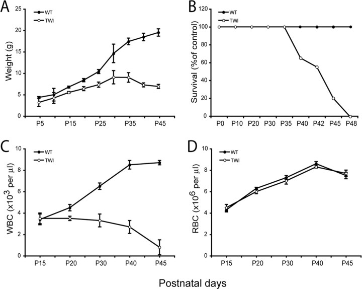
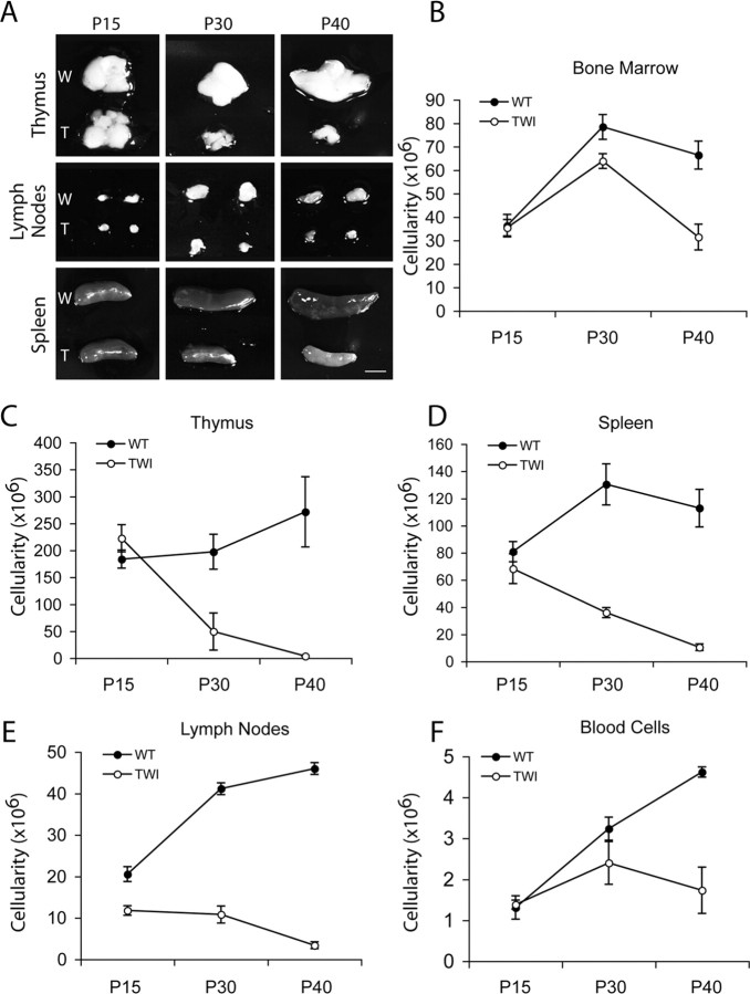
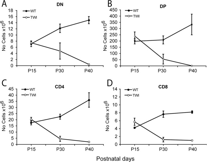
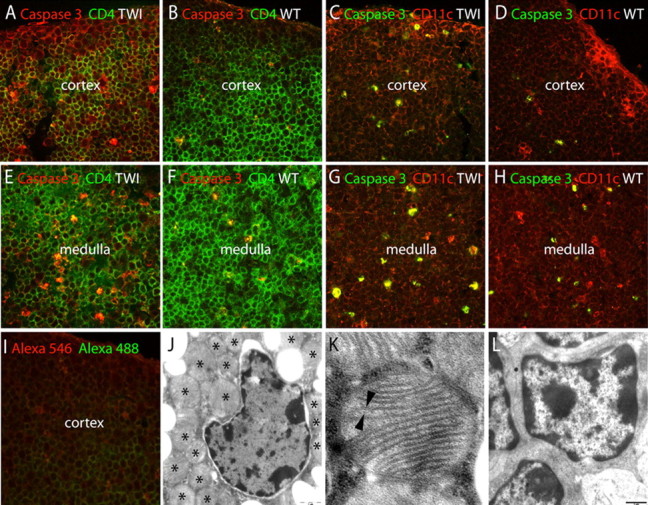
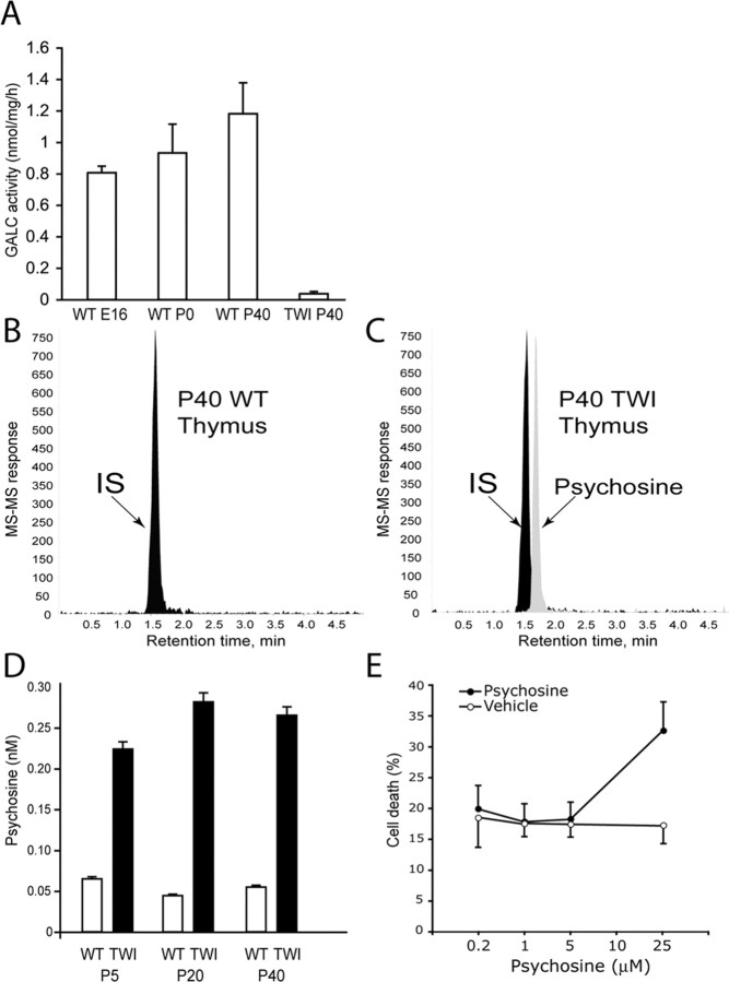
Lysosomal beta-galactosylceramidase deficiency results in demyelination and inflammation in the nervous system causing the neurological Krabbe disease. In the Twitcher mouse model of this disease, we found that neurological symptoms parallel progressive and severe lymphopenia. Although lymphopoiesis is normal before disease onset, primary and secondary lymphoid organs progressively degenerate afterward. This occurs despite preserved erythropoiesis and leads to severe peripheral lymphopenia caused by reduced numbers of T cell precursors and mature lymphocytes. Hematopoietic cell replacement experiments support the existence of an epigenetic factor in mutant mice reconcilable with a progressive loss of autonomic axons that hampers thymic functionality. We propose that degeneration of autonomic nerves leads to the irreversible thymic atrophy and loss of immune-competence. Our study describes a new aspect of Krabbe disease, placing patients at risk of immune-related pathologies, and identifies a novel target for therapeutic interventions.


Figures

Figure 1.
Figure 1.
GALC deficiency reduces circulating lymphocytes but not erythrocytes. A, B, Body weight and survival curves show that the deficiency of GALC prevents Twitcher mutant mice (TWI) to gain normal weight (A) and to survive beyond 48 d (B). C, D, Blood counts during postnatal development indicate that Twitcher mice develop a progressive lymphopenia (C), whereas red blood counts remain unaffected at all times (D). Data are expressed as the mean ± SE from 4–10 mice for each group per time point.
Figure 2.
Figure 2.
GALC deficiency is accompanied by atrophy of lymphoid organs. A, The pictures show a whole-mount comparison of primary and secondary lymphoid organs from Twitcher (T) and wild-type (W) littermate mice during postnatal age. Mutant mice showed a progressive atrophy of thymuses, lymph nodes, and spleens, which coincided in time with the appearance of neurological symptoms. B–F, Organ cellularity decreases during disease. Organ cellularity was defined by Trypan Blue negative cell counts of femurs- and tibiae-derived bone marrow cells (B); axillary, mesenteric, and inguinal pooled lymph nodes (E); 500 μl of peripheral blood (F); and intact thymus (C) and spleen (D). Twitcher mice (TWI) showed reduced cell numbers in the thymus, lymph nodes, and spleens between 15 and 30 d of age and remained lower than controls afterward. Bone marrow and blood cell numbers decreased in Twitcher mice starting from 30 d of age, indicating that these two compartments are affected during the late stage of the disease. Data are expressed as the mean ± SE from three to five mice for each group per time point.
Figure 3.
Figure 3.
GALC deficiency prevents normal thymocytes development. A–D, Thymocyte representation and distribution was evaluated by flow cytometry analysis of CD4, CD8 double negative (DN) (A), CD4, CD8 double positive (DP) (B), or CD4 (C) or CD8 (D) single positive cells. DN, DP, CD4, and CD8 thymocyte subsets decreased during disease progression in the Twitcher mice (TWI). Data are expressed as the mean ± SE from three to five mice for each group per time point.
Figure 4.
Figure 4.
Apoptosis and ultrastructural changes during GLD thymic atrophy. A–I, Apoptotic cell death was studied by activation of caspase 3 in Twitcher (TWI) and wild-type (WT) littermates at P15 (A, B, E, F) and P30 (C, D, G, H). Caspase 3-positive cells (red) were numerous in CD4 positive (green) T cells in the Twitcher thymic cortex (A) and medulla (E), respectively, controls. Caspase 3 was also active (green) in CD11c (red) stromal cells in the Twitcher thymic cortex (C) and medulla (G). I shows the background staining in P30 thymus. J–L, Transmission electron microscopy of ultrathin sections of Twitcher (J, K) and wild-type thymus (L) showed ultrastructural changes in thymic cells with abundant multilamellar giant bodies (J, asterisks), composed of bilayered membranes (K, arrowheads). Scale bar: (in J, L) 1 μm.
Figure 5.
Figure 5.
Determination of psychosine in GLD thymus. A, The GLD thymus lacks GALC activity. The enzyme was active in wild-type (WT) thymus from embryonic day 16 (E16) to P40. Data are expressed as the mean ± SE from three thymuses per time point. B, C, LC-MS-MS chromatograms showing the absence of psychosine in wild-type thymus (B) and the presence of the neurotoxin in Twitcher thymus (C). Lactosyl-sphingosine was used as an internal standard (IS). D, Quantification of psychosine during the postnatal development of the wild-type and Twitcher thymuses. Data are expressed as the mean ± SE from two to three thymuses for each time point per genotype. E, The effect of psychosine on thymocytes survival was studied in vitro using wild-type thymocytes from P15 mice. Cultures were exposed to different amounts of the sphingolipid for 1 d before mitochondrial membrane potential was determined as described. Data are expressed as the mean percentage of cell death ± SE from two cultures per dose.
Figure 6.
Figure 6.
Hematopoietic stem cell transplantation supports the existence of an epigenetic defect hampering thymic development in GLD mice. A, Transplantation of bone marrow-derived cells was performed between Twitcher (TWI) and wild-type congenic mice to test for epigenetic defects. Donor cell engraftment was measured in the blood at P40 and P75 and in the bone marrow (BM) at P75. Chimerism was similar in each of the three groups of transplanted mice, with an average engraftment of ∼75%. B, C, WBC counts were comparable in wild-type mice transplanted with either wild-type or Twitcher bone marrow cells but reduced in Twitcher mice transplanted with wild-type bone marrow. This suggests that the lymphopoiesis in the Twitcher mutant is affected by a noncell autonomous defect. D, E, Cellular composition of thymus (D) and spleen (E) after BM transplant. Normal numbers and distribution of thymocytes subsets were observed in mice that received wild-type or Twitcher bone marrow cells. In contrast, transplanted Twitcher mice showed a severe reduction in the number of DN, CD4, CD8, and DP thymic cells (D) and B220, CD4, CD8, and CD11b (E) splenocytes. Data are expressed as the mean ± SE from three to five mice for each group. *p < 0.01; **p < 0.05 versus wild-type recipient of wild-type transplanted cells.
Figure 7.
Figure 7.
Progressive axonal loss from autonomic fibers in the atrophied GLD thymus. A–D, Confocal immunohistochemistry of TH (green) showed reduced innervation of norepinephrine fibers in the Twitcher (TWI) cortex at P15 (A) and P30 (C), when compared with age-matched controls (B, D). A CD4 counterstaining (red) was used. E–H, Light microscopy immunohistochemistry for TH (E, F) and ChaT (G, H) in P40 thymuses showed reduced expression of TH (E) and ChaT (G) in the aged TWI thymus. I, The density of TH+ fibers was quantified and expressed as the number of TH+ fibers per 0.2 mm2 of thymic cortex at P20 and P30. Data averages two independent experiments. p < 0.05 versus wild type. J, Fold changes in expression of norepinephrine receptors (β1 and β2) was quantified by real-time PCR in RNA isolated from thymuses at P7 and P15. Data are normalized to the expression of the GAPDH housekeeping gene and compared with values obtained in age-matched wild-type tissues (the expression of which equals a value of 1). Data average two independent experiments. p < 0.05 versus wild type.

References

    1. Antonica A, Ayroldi E, Magni F, Paolocci N. Lymphocyte traffic changes induced by monolateral vagal denervation in mouse thymus and peripheral lymphoid organs. J Neuroimmunol. 1996;64:115–122. - PubMed
    1. Besedovsky H, Sorkin E. Network of immune-neuroendocrine interactions. Clin Exp Immunol. 1977;27:1–12. - PMC - PubMed
    1. Bhat S, Spitalnik SL, Gonzalez-Scarano F, Silberberg DH. Galactosyl ceramide or a derivative is an essential component of the neural receptor for human immunodeficiency virus type 1 envelope glycoprotein gp120. Proc Natl Acad Sci USA. 1991;88:7131–7134. - PMC - PubMed
    1. Brown DA, London E. Functions of lipid rafts in biological membranes. Annu Rev Cell Dev Biol. 1998;14:111–136. - PubMed
    1. Bullard W, Southard E. Difuse gliosis of the cerebral white matter in a child. J Nerv Ment Dis. 1906;33:188–193.

Publication types

MeSH terms