. 2007 Dec 12;27(50):13739-49.
doi: 10.1523/JNEUROSCI.3258-07.2007.

Tonic endovanilloid facilitation of glutamate release in brainstem descending antinociceptive pathways

Affiliations

Tonic endovanilloid facilitation of glutamate release in brainstem descending antinociceptive pathways

Katarzyna Starowicz et al. J Neurosci. .

Abstract

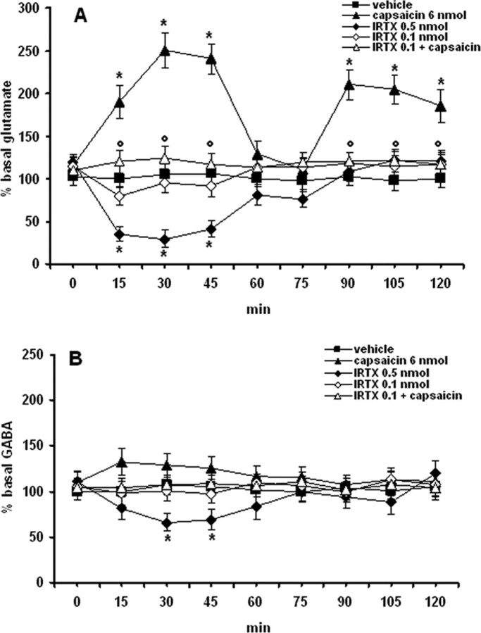
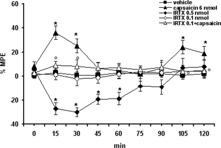
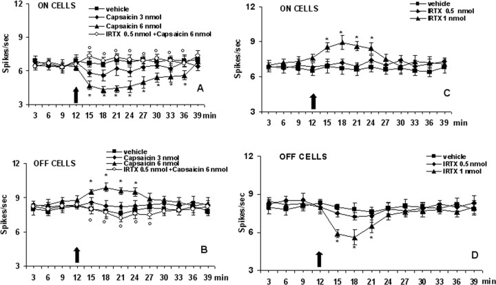
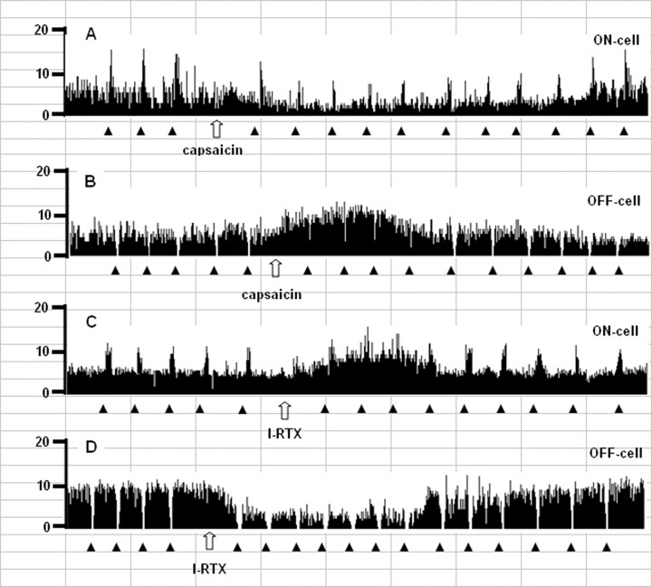
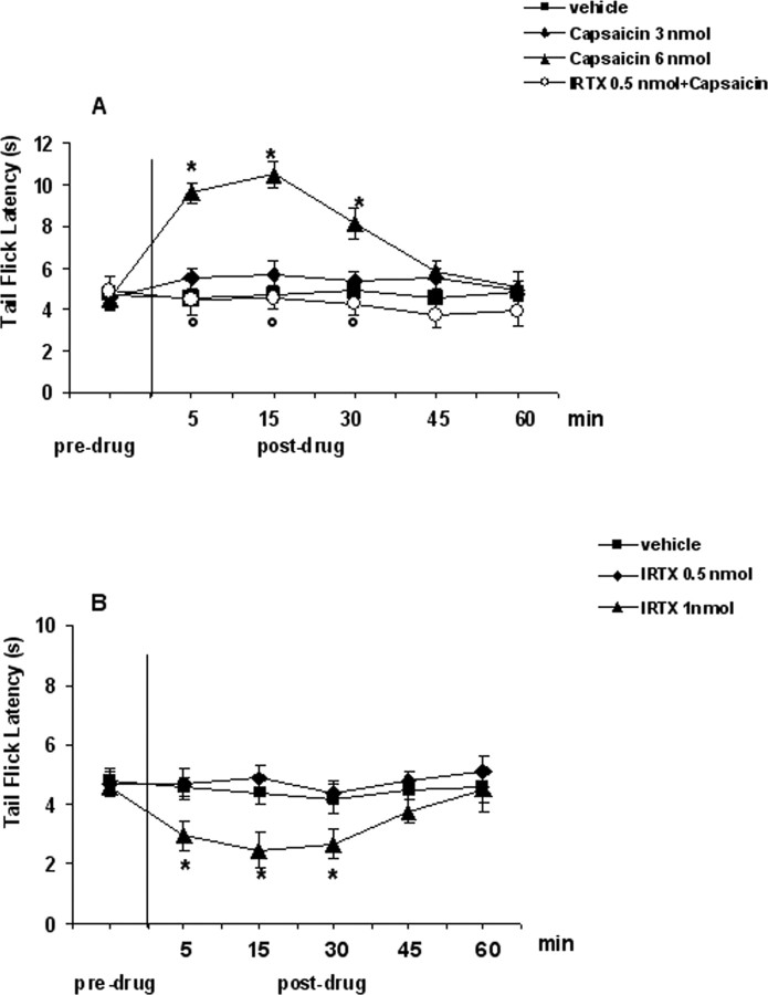
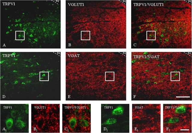
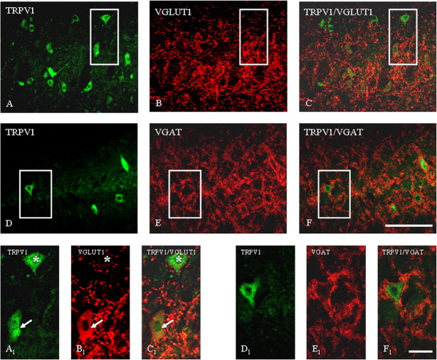
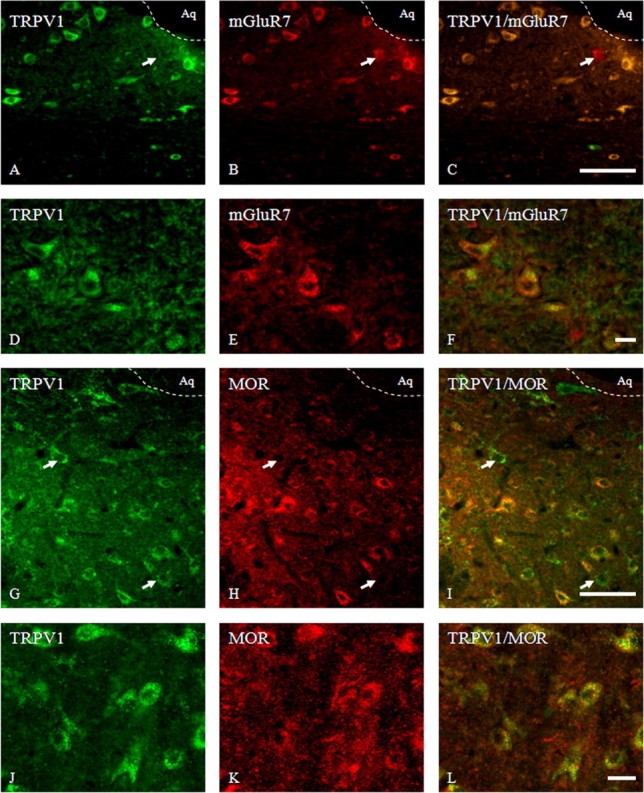
Activation of transient receptor potential vanilloid-1 (TRPV1) channels in the periaqueductal gray (PAG) activates OFF antinociceptive neurons of the rostral ventromedial medulla (RVM). We examined in rats the effect of intra-ventrolateral (VL)-PAG injections of TRPV1 agonists and antagonists on the nocifensive response to heat in the plantar test, neurotransmitter (glutamate and GABA) release in the RVM, and spontaneous and tail flick-related activities of RVM neurons. The localization of TRPV1 in VL-PAG and RVM neurons was examined using various markers of glutamatergic and GABAergic neurons. Intra-VL-PAG injection of capsaicin increased the threshold of thermal pain sensitivity, whereas the selective TRPV1 antagonist 5'-iodo-resiniferatoxin (I-RTX) facilitated nociceptive responses, and blocked capsaicin analgesic effect at a dose inactive per se. Intra-VL PAG capsaicin evoked a robust release of glutamate in RVM microdialysates. I-RTX, at a dose inactive per se, blocked the effect of capsaicin, and inhibited glutamate release at a higher dose. Antinociception and hyperalgesia induced by capsaicin and I-RTX, respectively, correlated with enhanced or reduced activity of RVM OFF cells. Immunohistochemical analyses suggested that several TRPV1-immunoreactive (ir) neurons in both the VL-PAG and RVM are glutamatergic and surrounded by glutamatergic and GABAergic terminals. Our data suggest that VL-PAG neurons respond to TRPV1 stimulation by releasing glutamate into the RVM, thereby activating OFF cells and producing analgesia. The results obtained with the TRPV1 antagonist alone suggest that this pathway is tonically activated by endovanilloids.


Figures

Figure 1.
Figure 1.
Effects of intraventrolateral PAG injection of vehicle, capsaicin (6 nmol/rat), I-RTX (0.1 and 0.5 nmol/rat), and capsaicin (6 nmol/rat) in combination with I-RTX (0.1 nmol/rat) on RVM dialysate levels of glutamate (A) and GABA (B). Each point represents the mean ± SEM of amino acid extracellular concentrations as a percentage of the basal values (8–10 rats per group). * indicates significant difference versus vehicle; ° indicates significant difference versus capsaicin (6 nmol/rat). p values <0.05 were considered statistically significant.
Figure 2.
Figure 2.
Thermal nociception was evaluated by using a plantar test and expressed as percentage of the maximum possible effect: %MPE = [(test latency) − (control latency)/(cutoff time) − (control latency)] × 100. Vehicle (10% DMSO in ACSF), capsaicin (6 nmol/rat), I-RTX (0.1 and 0.5 nmol/rat), and capsaicin (6 nmol/rat) in combination with I-RTX (0.1 nmol/rat) were microinjected into the ventrolateral PAG. Nociceptive responses were measured every 15 min for a period of 2 h. Each data point represents the mean ± SEM of 8–10 animals per group. * indicates significant differences versus vehicle; °, indicates significant differences versus capsaicin (6 nmol/rat). p values <0.05 were considered statistically significant.
Figure 3.
Figure 3.
Schematic illustration of the location of PAG microinjection sites (A), RVM ON- or OFF-cell recording sites (B), and RVM microdialysis probes location (C). Vehicle or drug microinjections were performed into the ventrolateral PAG matter (filled circles) (A) while cell recording was performed by lowering a tungsten electrode into the RVM (B). Filled triangles represent ON-cells and open triangles the OFF-cell sites. The location of microdialysis probe (C) was also histologically confirmed and marked as correct (black bars) when the probe tip fell within the RVM areas here studied. Many sites are not shown because of overlap of symbols. Distances from the interaural line are indicated.
Figure 4.
Figure 4.
Effect of capsaicin and 5′-Iodo-resiniferatoxin (I-RTX) on the spontaneous firing of RVM ON (A, C) or OFF (B, D) cells. A and B show the effect of intraventrolateral PAG administration of vehicle, capsaicin (3 and 6 nmol/rat) alone, or capsaicin (6 nmol/rat) in combination with I-RTX (0.5 nmol/rat). C and D shown the effect of I-RTX (0.5 and 1 nmol/rat) alone. The black arrow indicates drug microinjections. Each point represents the mean ± SEM of 8–10 neurons. * indicates significant differences versus vehicle; ° indicates significant differences versus capsaicin (6 nmol/rat). p values <0.05 were considered statistically significant.
Figure 5.
Figure 5.
Examples of ratemeter records that illustrate the effects of intra-PAG microinjections of capsaicin (6 nmol/rat) (A, B) or I-RTX (1 nmol/rat) (C, D) on either the ongoing or tail flick-related discharges of identified RVM ON (A, C) and OFF (B, D) cells. Traces report overall firing before and after drug injections into the ventrolateral PAG. Filled triangles indicate tail flick trials, 1 s bins. Open arrows show the time of microinjections within the ventrolateral PAG. Time units, 1 cell = 7 min.
Figure 6.
Figure 6.
Tail flick latencies before and after microinjections into the ventrolateral PAG of vehicle, capsaicin (3 and 6 nmol/rat) alone or in combination with I-RTX (0.5 nmol/rat) (A), and vehicle or I-RTX (0.5 and 1 nmol/rat) (B). Each point represents the mean ± SEM of 8–10 observations. * indicates significant differences versus vehicle; ° indicates significant differences versus capsaicin (6 nmol/rat). p values <0.05 were considered statistically significant.
Figure 7.
Figure 7.
Immunohistochemical localization of TRPV1 receptors and VGLUT1 (A–C) and TRPV1 receptors and VGAT (D–F) in rat ventrolateral PAG as determined by the immunofluorescence technique. A–C, General view of the immunoreactive distribution in the ventrolateral subregion of the PAG of TRPV1 (A), VGLUT1 (B), and TRPV1/VGLUT1 (merged image) (C). A1–C1, High magnification of respective boxed areas of A–C. Note the dense TRPV1 receptor immunolabeling in cellular cytoplasm and processes (in green) and nerve terminals labeled for VGLUT1 (in red). D–F, General view of the immunoreactive distribution in the ventrolateral subregion of the PAG of TRPV1 (D), VGAT (E), and TRPV1/VGAT (merged image) (F). D1–F1, High magnification of respective boxed areas of D–F. Note the dense TRPV1 receptor immunolabeling in cytoplasm on cell bodies and neuronal processes (in green) and terminals labeled for VGAT (in red). Aq, Lumen of aqueduct. Scale bars: A–F, 80 μm; A1–F1, 20 μm. Images are representative of those obtained in nine sections per rat.
Figure 8.
Figure 8.
Micrographs demonstrating fluorescent staining of neurons in the RVM for TRPV1 receptor and VGLUT1 (A–C) and for TRPV1 receptor and VGAT (D–F). A–C, Overview of the RVM region examined for TRPV1 and VGLUT1 localization; solid line box marks the region shown in A1–C1. Note that some TRPV1-labeled cells are surrounded by VGLUT1-positive nerve terminals (asterisk), whereas other cells are positive for both TRPV1 and VGLUT1 (arrows). D–F, Note constant TRPV1 labeling of cell membranes and cytoplasm in the RVM (D, D1) and VGAT-positive terminals (E, E1) surrounding TRPV1-positive cells (F, F1). Scale bar: A–F, 60 μm; A1–F1, 15 μm. Images are representative of those obtained in nine sections per rat.
Figure 9.
Figure 9.
Photomicrographs of coronal sections through the ventrolateral PAG showing the same area immunolabeled for TRPV1 in green, mGluR7 or μ opioid receptor (MOR) in red, and both in yellow in the merged images; and in the RVM representing neurons marked for TRPV1 (green) and mGluR7 (red) and merged images (yellow). A–C, Double immunofluorescence for TRPV1 and mGluR7 in the ventrolateral PAG. Note colocalization of TRPV1 and mGluR7 in the vast majority of neurons; just a few cells were only mGluR7 positive (arrow). D–F, Intense cytoplasmic localization for TRPV1 and mGluR7 on cell bodies of RVM neurons. G–I, Double immunofluorescence for TRPV1 and MOR in the ventrolateral PAG region showing TRPV1 distribution with postsynaptic MOR (yellow cells) in the majority of neurons stained, although some cells were also simply TRPV1-ir (arrows). J–L, Strong cytoplasmic colocalization for TRPV1 and MOR in a vast majority of cell bodies of RVM neurons. Aq, Lumen of aqueduct. Scale bars: A–C, G–I, 80 μm; D–F, J–L, 15 μm. Images are representative of those obtained in nine sections per rat.

Similar articles

Cited by

References

    1. Azkue JJ, Mateos JM, Elezgarai I, Benitez R, Lazaro E, Streit P, Grandes P. Glutamate-like immunoreactivity in ascending spinofugal afferents to the rat periaqueductal grey. Brain Res. 1998;790:74–81. - PubMed
    1. Basbaum AI, Fields HL. Endogenous pain control systems: brainstem spinal pathways and endorphin circuitry. Annu Rev Neurosci. 1984;7:309–338. - PubMed
    1. Behbehani MM. Functional characteristics of the midbrain periaqueductal gray. Prog Neurobiol. 1995;46:575–605. - PubMed
    1. Behbehani MM, Fields HL. Evidence that an excitatory connection between the periaqueductal gray and nucleus raphe magnus mediates stimulation produced analgesia. Brain Res. 1979;170:85–93. - PubMed
    1. Benninger F, Freund TF, Hajos N. Control of excitatory synaptic transmission by capsaicin is unaltered in TRPV(1) vanilloid receptor knockout mice. Neurochem Int. 2007 in press. - PMC - PubMed

MeSH terms