

Neuronal activity related to reward size and rewarded target position in primate supplementary eye field
- PMID: 18077686
- PMCID: PMC6673633
- DOI: 10.1523/JNEUROSCI.2693-07.2007
Neuronal activity related to reward size and rewarded target position in primate supplementary eye field
Abstract
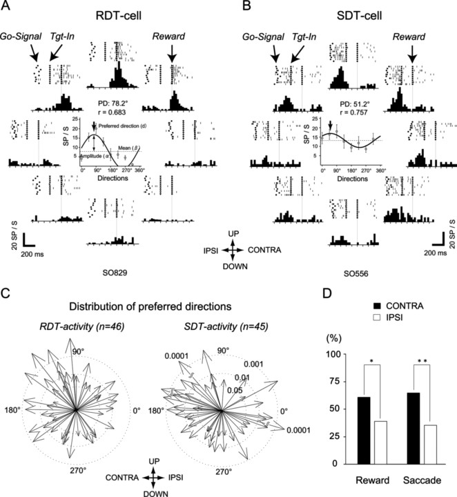
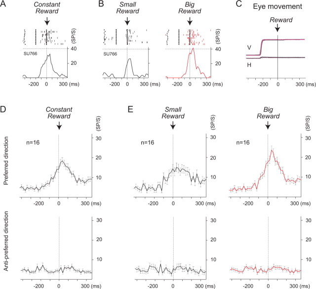
Several areas of the macaque brain are known to be related to the reward during the performance of saccadic eye-movement tasks. Neurons in the supplementary eye field (SEF) have been reported to be involved in the prediction and detection of a reward. We describe a group of neurons in the SEF that became active during the period of reward delivery after saccades toward a specific direction, but showed weaker activity in other directions, although the same amount of reward was given in each direction. Moreover, this directional reward activity was modulated by the reward size. Our results demonstrate that the SEF cells may reflect both reward amount and target positions toward which a movement was rewarded, and suggest that they may play an important role in providing information about the value of each saccade according to the spatial target location.
Figures
Similar articles
-
Neurons in the supplementary eye field of rhesus monkeys code visual targets and saccadic eye movements in an oculocentric coordinate system.J Neurophysiol. 1996 Aug;76(2):825-48. doi: 10.1152/jn.1996.76.2.825. J Neurophysiol. 1996. PMID: 8871203
-
The dorsomedial frontal cortex of the macaca monkey: fixation and saccade-related activity.Exp Brain Res. 1992;89(3):571-80. doi: 10.1007/BF00229882. Exp Brain Res. 1992. PMID: 1644122
-
Supplementary eye field: representation of saccades and relationship between neural response fields and elicited eye movements.J Neurophysiol. 2000 Nov;84(5):2605-21. doi: 10.1152/jn.2000.84.5.2605. J Neurophysiol. 2000. PMID: 11068002
-
Supplementary eye field encodes option and action value for saccades with variable reward.J Neurophysiol. 2010 Nov;104(5):2634-53. doi: 10.1152/jn.00430.2010. Epub 2010 Aug 25. J Neurophysiol. 2010. PMID: 20739596 Free PMC article.
-
Cellular activity in the supplementary eye field during sequential performance of multiple saccades.J Neurophysiol. 2002 Dec;88(6):3541-5. doi: 10.1152/jn.00299.2002. J Neurophysiol. 2002. PMID: 12466467
Cited by
-
Alterations in the functional neural circuitry supporting flexible choice behavior in autism spectrum disorders.Transl Psychiatry. 2016 Oct 11;6(10):e916. doi: 10.1038/tp.2016.161. Transl Psychiatry. 2016. PMID: 27727243 Free PMC article.
-
A role for the ventral premotor cortex beyond performance monitoring.Proc Natl Acad Sci U S A. 2009 Nov 3;106(44):18815-9. doi: 10.1073/pnas.0910524106. Epub 2009 Oct 21. Proc Natl Acad Sci U S A. 2009. PMID: 19846763 Free PMC article.
-
Decision-making, behavioral supervision and learning: an executive role for the ventral premotor cortex?Neurotox Res. 2010 Nov;18(3-4):416-27. doi: 10.1007/s12640-010-9194-y. Epub 2010 Apr 20. Neurotox Res. 2010. PMID: 20405351 Review.
-
Role of supplementary eye field in saccade initiation: executive, not direct, control.J Neurophysiol. 2010 Feb;103(2):801-16. doi: 10.1152/jn.00221.2009. Epub 2009 Nov 25. J Neurophysiol. 2010. PMID: 19939963 Free PMC article.
-
Neuronal Correlates of Serial Decision-Making in the Supplementary Eye Field.J Neurosci. 2018 Aug 15;38(33):7280-7292. doi: 10.1523/JNEUROSCI.3643-17.2018. Epub 2018 Jul 16. J Neurosci. 2018. PMID: 30012690 Free PMC article.
References
-
- Amador N, Schlag-Rey M, Schlag J. Reward-predicting and reward-detecting neuronal activity in the primate supplementary eye field. J Neurophysiol. 2000;84:2166–2170. - PubMed
-
- Amador N, Schlag-Rey M, Schlag J. Primate antisaccade. II. Supplementary eye field neuronal activity predicts correct performance. J Neurophysiol. 2004;91:1672–1689. - PubMed
-
- Chen LL, Wise SP. Supplementary eye field contrasted with the frontal eye field during acquisition of conditional oculomotor associations. J Neurophysiol. 1995;73:1122–1134. - PubMed
-
- Chen LL, Wise SP. Conditional oculomotor learning: population vectors in the supplementary eye field. J Neurophysiol. 1997;78:1166–1169. - PubMed
Publication types
MeSH terms
LinkOut - more resources
Full Text Sources