. 2007 Dec 12;27(50):13793-801.
doi: 10.1523/JNEUROSCI.3489-07.2007.

Electrical stimulation of spared corticospinal axons augments connections with ipsilateral spinal motor circuits after injury

Affiliations

Electrical stimulation of spared corticospinal axons augments connections with ipsilateral spinal motor circuits after injury

Marcel Brus-Ramer et al. J Neurosci. .

Abstract

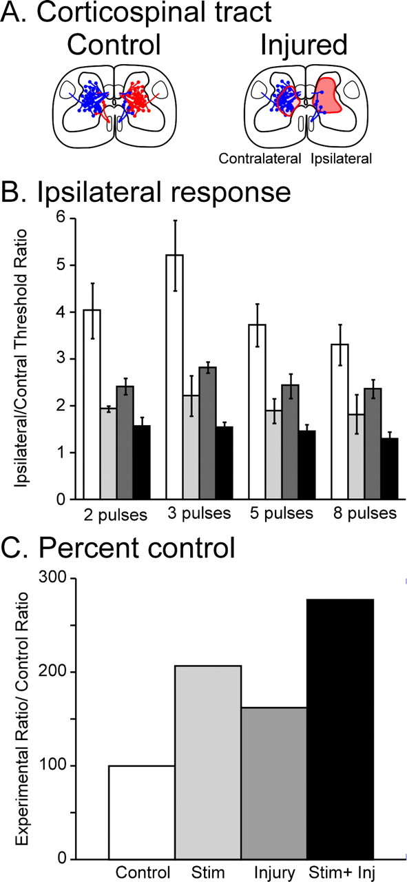
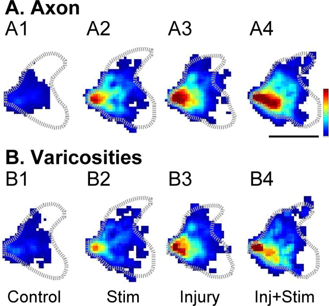
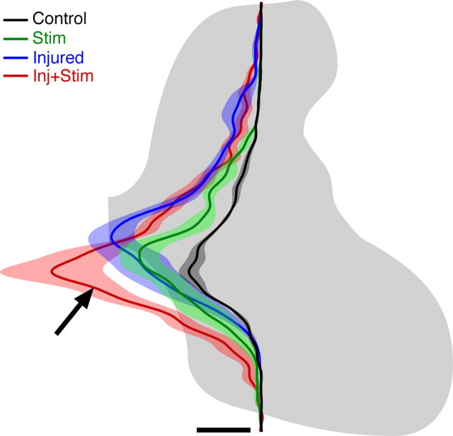
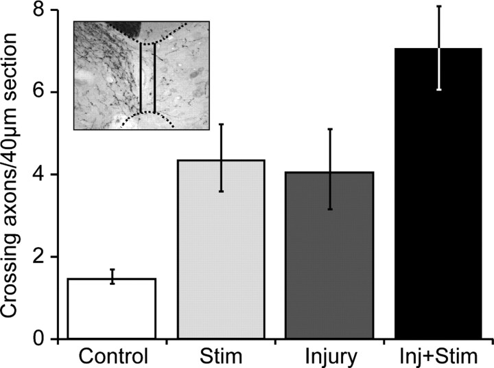
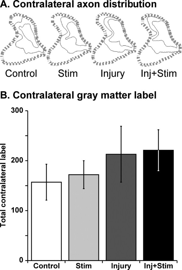
Activity-dependent competition shapes corticospinal (CS) axon outgrowth in the spinal cord during development. An important question in neural repair is whether activity can be used to promote outgrowth of CS axons in maturity. After injury, spared CS axons sprout and make new connections, but often not enough to restore function. We propose that electrically stimulating spared axons after injury will enhance sprouting and strengthen connections with spinal motor circuits. To study the effects of activity, we electrically stimulated CS tract axons in the medullary pyramid. To study the effects of injury, one pyramid was lesioned. We studied sparse ipsilateral CS projections of the intact pyramid as a model of the sparse connections preserved after CNS injury. We determined the capacity of CS axons to activate ipsilateral spinal motor circuits and traced their spinal projections. To understand the separate and combined contributions of injury and activity, we examined animals receiving stimulation only, injury only, and injury plus stimulation. Both stimulation and injury alone strengthened CS connectivity and increased outgrowth into the ipsilateral gray matter. Stimulation of spared axons after injury promoted outgrowth that reflected the sum of effects attributable to activity and injury alone. CS terminations were densest within the ventral motor territories of the cord, and connections in these animals were significantly stronger than after injury alone, indicating that activity augments injury-induced plasticity. We demonstrate that activity promotes plasticity in the mature CS system and that the interplay between activity and injury preferentially promotes connections with ventral spinal motor circuits.


Figures

Figure 1.
Figure 1.
Chronic PT stimulation or injury alone increase the strength of ipsilateral connections, which were further strengthened by combined injury and stimulation. A, Schematic model of CS termination field in the normal cervical spinal cord (Control) and the changes in termination density after PT lesion (Injured). Blue axons derive from the intact PT; red axons derive from the injured PT; pink shading in injured CS shows the former position of the lesioned axons. B, Ratios of ipsilateral current threshold to contralateral current threshold (Ipsilateral/Contral Threshold Ratio) needed to evoke responses in the deep branch of the radial nerve to PT stimulus trains of two, three, five, and eight pulses. A lower value in the ipsilateral–contralateral ratio indicates augmented access to ipsilateral spinal motor circuits. C, Summary graph showing percentage change in ipsilateral–contralateral threshold ratio. The values for each group were calculated from an average threshold response to all stimulus train lengths. Stimulation (Stim) and injury alone both show increased enhanced capacity to evoke ipsilateral motor responses, which is augmented further in the combined condition (Stim+Inj).
Figure 2.
Figure 2.
Stimulation and injury each promote increased density of ipsilateral CS terminations, which is further augmented in the combined condition. A, Axon density (terminals and preterminal axons) maps. B, Bouton density maps. Color coded average density maps are shown for controls (A1, B1; n = 4), animals with PT stimulation alone (Stim; A2, B2; n = 4), animals with injury (PTx) alone (A3, B3; n = 5), and animals with both stimulation and injury (Inj+Stim; A4, B4; n = 5). The ipsilateral gray matter border (gray hatched line) shows averaged borders from the individual animals in the group. Calibration: 500 μm; Color scale: A, 0–5.7 μm axon/μm2 area; B, 0–0.7 boutons/μm2 area.
Figure 3.
Figure 3.
Stimulation (Stim) and injury alone and in combination (Inj+Stim) augment total ipsilateral CS termination axon length. A, Total average ipsilateral axon length in the controls, stimulation alone, injury alone, and combined injury and stimulation. Stimulation (p = 0.011) and injury (p = 0.003) alone each augmented total axon length significantly compared with control. Combined, there was a larger increase (p = 0.002). p values were calculated from t test with Bonferroni/Dunn correction. B, Ventral ipsilateral axon length in the controls, stimulation alone, injury alone, and combined condition (ANOVA, F = 7.37; p = 003). Combined stimulation and injury significantly augmented axon length in this region compared with injury alone (t test; p = 0.04).
Figure 4.
Figure 4.
Stimulation after injury shifts the distribution of CS terminations ventrally in the gray matter. Dorsoventral distributions of mean axon density from the dorsal to the ventral borders of the ipsilateral gray mater are shown. Each line plots the average density of axon terminations (preterminal and terminal axons; no boutons) of the animals in each group; light shading plots ± SEM. Data from controls are shown in black (n = 4 rats); stimulation alone (Stim; n = 4), green; injury alone (n = 5), blue; and combined injury and stimulation (Inj+Stim; n = 5), red. The arrow indicates a ventral location where axon length is greatest in the injury and stimulation group. To normalize for differences in the size of the gray matter across animals, all graphs were interpolated to 1000 points between the dorsal and ventral margins of the gray matter. Calibration: 30 μm of labeled axons.
Figure 5.
Figure 5.
Stimulation and injury alone and combination augment the number of axons crossing in the cervical spinal gray matter. Crossing axon counts per 40-μm-thick transverse section for control, stimulation (Stim), injury, and combined stimulation and injury (Inj+Stim). There was a significant difference in lamina 10 axons between groups (ANOVA, F = 6.83; p = 0.005), and importantly, combined stimulation and injury significantly augmented crossing axons compared with injury alone (Bonferroni/Dunn post hoc, p = 0.0005). Inset, Representative example of axons in lamina 10 of the gray matter. Solid vertical lines indicate the midline (left) and 25 μm ipsilateral to midline (right), and dashed lines mark the dorsal and ventral gray matter borders. Only axons within the 25 μm region were counted.
Figure 6.
Figure 6.
Contralateral CS terminations do not respond significantly to stimulation (Stim) and injury alone and in combination (Inj+Stim). A, Distributions of regions of densest CS labeling (inner solid line; 35%) and total labeling (outer solid line; total label). Axon label was determined by optical density after reducing background signal to zero. Data from the four animal groups are shown: controls (n = 4), stimulated alone (n = 4), injury alone (n = 5), and combined injury and stimulation (n = 5). B, Total contralateral label for each animal group. There were no significant differences between groups (ANOVA, F = 0.56; p = 0.655).

Comment in

References

    1. Alstermark B, Ogawa J, Isa T. Lack of monosynaptic corticomotoneuronal EPSPs in rats: disynaptic EPSPs mediated via reticulospinal neurons and polysynaptic EPSPs via segmental interneurons. J Neurophysiol. 2004;91:1832–1839. - PubMed
    1. Aoki M, Fujito Y, Satomi H, Kurosawa Y, Kasaba T. The possible role of collateral sprouting in the functional restitution of corticospinal connections after spinal hemisection. Neurosci Res. 1986;3:617–627. - PubMed
    1. Bareyre FM, Kerschensteiner M, Raineteau O, Mettenleiter TC, Weinmann O, Schwab ME. The injured spinal cord spontaneously forms a new intraspinal circuit in adult rats. Nat Neurosci. 2004;7:269–277. - PubMed
    1. Bradbury EJ, Moon LD, Popat RJ, King VR, Bennett GS, Patel PN, Fawcett JW, McMahon SB. Chondroitinase ABC promotes functional recovery after spinal cord injury. Nature. 2002;416:636–640. - PubMed
    1. Brosamle C, Schwab ME. Cells of origin, course, and termination patterns of the ventral, uncrossed component of the mature rat corticospinal tract. J Comp Neurol. 1997;386:293–303. - PubMed

Publication types