. 2007 Dec 12;27(50):13866-74.
doi: 10.1523/JNEUROSCI.3136-07.2007.

A mutation in the cone-specific pde6 gene causes rapid cone photoreceptor degeneration in zebrafish

Affiliations

A mutation in the cone-specific pde6 gene causes rapid cone photoreceptor degeneration in zebrafish

George Stearns et al. J Neurosci. .

Abstract

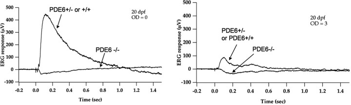
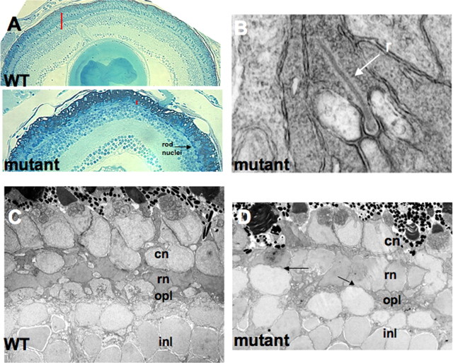
Photoreceptor degeneration is a common cause of inherited blindness worldwide. We have identified a blind zebrafish mutant with rapid degeneration of cone photoreceptors caused by a mutation in the cone phosphodiesterase c (pde6c) gene, a key regulatory component in cone phototransduction. Some rods also degenerate, primarily in areas with a low density of rods. Rod photoreceptors in areas of the retina that always have a high density of rods are protected from degeneration. Our findings demonstrate that, analogous to what happens to rod photoreceptors in the rd1 mouse model, loss of cone phosphodiesterase leads to rapid degeneration of cone photoreceptors. Furthermore, we propose that cell density plays a key role in determining whether rod photoreceptors degenerate as a secondary consequence to cone degeneration. Our zebrafish mutant serves as a model for developing therapeutic treatments for photoreceptor degeneration in humans.


Figures

Figure 1.
Figure 1.
External morphology and retinal phenotype of PDE6c −/− through 7 dpf. Dorsal (A) and lateral (B) views of pde6c w59 and sibling nonblind fish. Notice the normal eye size and development of a swim bladder (asterisk in B) in the mutant. The mutation is recessive and one-quarter of the larvae from a mating cross between two heterozygous fish are blind at 5 dpf. pde6c w59 retinas at 3 dpf (C), 4 dpf (D), and 7 dpf (E). C, At day 3, cone outer segments are apparent in the mutant retina. The arrow points to an intact cone outer segment. D, By day 4, cones in the central one-half of the retina have degenerated. The bottom right panel labeled “rods” points to the elongated rod outer segments in the ventral patch of the retina. E, At 7 dpf in WT retinas, cone outer segments dominate the outer nuclear layer. In mutants, cones are markedly absent, and rods are distributed throughout the retina and their outer segments appear aggregated (red oval). F, Whole-mount staining of rod photoreceptors at 4 dpf. The image was collected at the level of photoreceptor outer segments. The overall distribution of rods across the retina appears similar in mutant and WT eyes. Rod outer segments are pronounced in the mutant. The antibody used (4c12) labels an unknown epitope specific to rod photoreceptors. Please see text for more details.
Figure 2.
Figure 2.
Sequence comparison between pde6c w59 fish and WT fish and cone-specific expression of pde6c. A, The mutation is in a splice acceptor site between exon 11 and exon 12. The red arrow points to the mutation (A to G) and the black arrow marks the beginning of exon 12. Mutant sequence is on top, and WT sequence is on the bottom. B–E show in situ hybridization on WT zebrafish larvae using pde6c antisense RNA. B, At 53 hpf, staining was only detected in the pineal (arrow). C, D, At 72 hpf, staining in the ventral patch (arrow) and ventral anterior portion (leftward arrow) of the eye was detected. E, Robust symmetric staining across the ONL was evident at 5 dpf.
Figure 3.
Figure 3.
ERGs recorded from whole zebrafish larvae at 20 dpf. Note the heterozygous and WT fish show a much larger b-wave response in bright light [left panel optical density filter (OD) = 0.0] than in dim light (right panel OD = 3.0). Only a possible small a-wave was noted in the mutant larvae.
Figure 4.
Figure 4.
Histology of WT and pde6c w59 retinas at 8 dpf. A and C show Fret43 labeling for double cones. B and D show 4c12 labeling for rods. Red, Antibody staining; blue, DAPI staining for nuclei; green, nyx::MYFP for bipolar cells (bp) (Schroeter et al., 2006). In the WT retina, the cones (A) are evenly distributed throughout the outer nuclear layer. The rods (B) are less dense and their distribution is asymmetric but they also are found throughout the photoreceptor layer. In the pde6c w59 fish, cones (C) and rods (D) are localized to the retinal periphery. Note the absence of both rods and cones (arrows in C and D) near the center of the retina, around the optic nerve. E–H show high-magnification bipolar cell morphology in the WT and pde6c w59. The arrows in F point to displaced bipolar nuclei and axonal processes in pde6c w59. G and H show a tangential section of bipolar dendrites at the level of the photoreceptor synapse. Note the dramatic reduction in number of dendritic processes and the lack of a regular pattern in the mutant compared with WT.
Figure 5.
Figure 5.
Histology of WT and pde6c w59 retinas at 6 weeks. A compares light micrographs of WT and mutant retinas. Note the proximity of the OPL to the pigmented epithelium (red line) in mutant compared with WT. The arrow in A points to rod nuclei. B shows a morphologically normal rod spherule (r) from the pde6c w59 retinal periphery. C and D compare the OPL of WT and pde6c w59 central retinas. The arrows in D highlight regions in the mutant where the OPL is missing. cn, Cone nuclei; rn, rod nuclei.
Figure 6.
Figure 6.
Rod versus cone distribution in 3 month pde6c w59 mutant retinas. A and C show Fret43 labeling for double cones. B and D show 4c12 labeling for rods. Red, Antibody staining; blue, DAPI staining for nuclei. The central retina is shown in A and B, whereas C and D show staining within the retinal margin. Note the absence of most Fret43 (i.e., cone) labeling in the central retina (arrow in A) but a regular palisade of cones near the margin (C). This is consistent with continual generation of cones occurring at the retinal margin throughout the life of the fish. In contrast, the rods have long outer segments both at the margin and toward the central retina (D, B, respectively). E and F show a comparison of rod spherules in WT versus pde6c w59 retinas at 3 months using confocal microscopy. The retinas were stained with the rod-specific antibody 4c12. The white arrow in each panel points to a group of three spherules. The red-stained spherule surrounds a black (unstained) region that is the invaginating synapse. No obvious differences were noted between mutant and WT. See Results for cell counts.
Figure 7.
Figure 7.
Schematic of cell loss and repopulation in the zebrafish pde6c w59 mutant. At 3 dpf, cones are evenly distributed throughout the ONL and rods are less dense and asymmetrically distributed. Between 3 and 4 dpf, cones die. Subsequently, rods degenerate in the central retina. Cell growth at the retinal margins (and possibly also the within the INL and ONL) continues to generate new rods and cones. The cones continue to die because of the pde6c w59 allele. The rod photoreceptors eventually repopulate the mutant retina.

References

    1. Alexander JJ, Umino Y, Everhart D, Chang B, Min SH, Li Q, Timmers AM, Hawes NL, Pang JJ, Barlow RB, Hauswirth WW. Restoration of cone vision in a mouse model of achromatopsia. Nat Med. 2007;13:685–687. - PMC - PubMed
    1. Bernardos RL, Barthel LK, Meyers JR, Raymond PA. Late-stage neuronal progenitors in the retina are radial Müller glia that function as retinal stem cells. J Neurosci. 2007;27:7028–7040. - PMC - PubMed
    1. Biel M, Seeliger M, Pfeifer A, Kohler K, Gerstner A, Ludwig A, Jaissle G, Fauser S, Zrenner E, Hofmann F. Selective loss of cone function in mice lacking the cyclic nucleotidegated channel CNG3. Proc Natl Acad Sci USA. 1999;96:7553–7557. - PMC - PubMed
    1. Bilotta J, Saszik S, Sutherland SE. Rod contributions to the electroretinogram of the dark-adapted developing zebrafish. Dev Dyn. 2001;222:564–570. - PubMed
    1. Bowes C, Li T, Danciger M, Baxter LC, Applebury ML, Farber DB. Retinal degeneration in the rd mouse is caused by a defect in the beta subunit of rod cGMP-phosphodiesterase. Nature. 1990;347:677–680. - PubMed

Publication types

MeSH terms

Substances