. 2008 Mar;82(5):2241-9.
doi: 10.1128/JVI.02093-07. Epub 2007 Dec 12.

Hepatitis C virus genotype 1a growth and induction of autophagy

Affiliations

Hepatitis C virus genotype 1a growth and induction of autophagy

Malika Ait-Goughoulte et al. J Virol. 2008 Mar.

Erratum in

  • J Virol. 2008 Jul;82(13):6783

Abstract

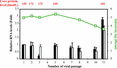
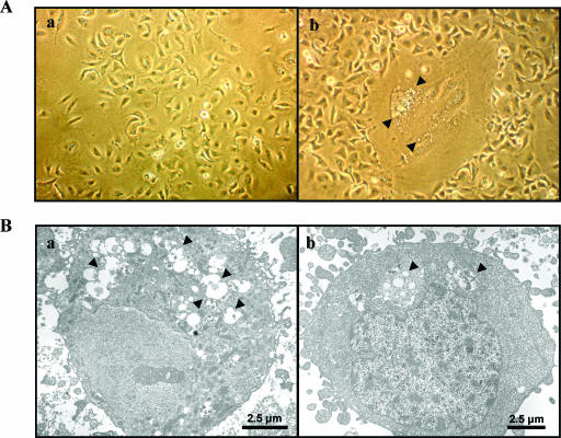
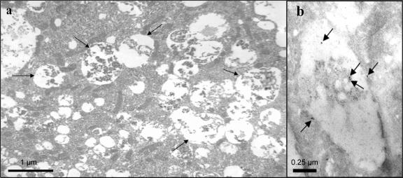
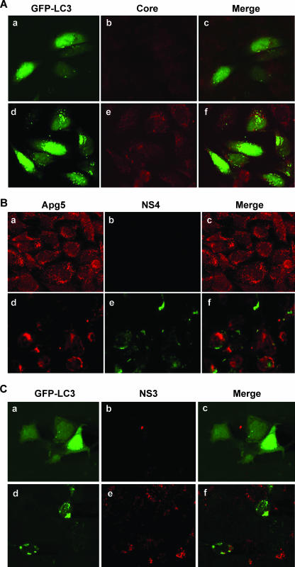
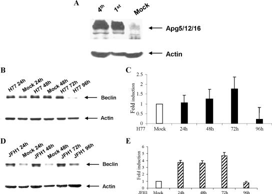
We have previously reported that immortalized human hepatocytes (IHH) support the generation of infectious hepatitis C virus (HCV) genotype 1a (clone H77). In the present study, we have investigated the growth of HCV genotype 1a (clone H77) through serial passages and accompanying changes in IHH in response to infection. Eleven serial passages of HCV genotype 1a (clone H77) in IHH were completed. Virus replication was ascertained from the presence of HCV-specific sequences, the detection of core antigen, the virus genome copy number, and the virus titer in IHH culture fluid. Electron microscopy suggested that HCV infection induces autophagic vacuole formation in IHH. Fluorescence microscopy displayed localization of autophagic markers, microtubule-associated protein-1 light chain-3 and Apg5, on the vacuoles of HCV-infected hepatocytes. Taken together, our results suggested that HCV genotype 1a (clone H77) can be serially passaged in IHH and that HCV infection induces an autophagic response in hepatocytes.


Figures

FIG. 1.
FIG. 1.
Replication of HCV genotype 1a (clone H77) after serial passage in IHH. The HCV replication and secretion of virus in culture medium were examined using different parameters. The relative copy number of HCV RNA at the intracellular level and in culture supernatant were measured by real-time PCR and calculated as described in Materials and Methods. The results are presented with standard errors from three independent experiments. Mock-infected or HCV-infected IHH culture supernatant was tested for core protein detection by ELISA. HCV titer (ffu/ml) was determined by the immunofluorescence of IHH 3 days after infection with filtered culture medium for the detection of NS4 protein expression. Relative HCV RNA in cell culture fluid (black bars) and HCV GAPDH ratio in cellular RNA (white bars) are shown. Infectivity (ffu/ml) is shown as a green line, and core protein level is shown on top.
FIG. 2.
FIG. 2.
Cellular changes following HCV genotype 1a (clone H77) infection of IHH. (A) Comparison of the cellular changes, by using light microscopy, between the uninfected (panel a) and the HCV-infected (panel b) IHH. Arrowheads in the photomicrograph indicate vacuole-like inclusions in larger cells. (B) Cellular changes were also compared by electron microscopy of uninfected (panel a) and HCV-infected (panel b) IHH. Numerous autophagic vacuoles were observed in the virus-infected cells compared to the uninfected cells and are indicated by arrows.
FIG. 3.
FIG. 3.
Electron micrographs showing cytoplasmic contents in autophagic vacuoles. IHH were infected with HCV genotype 1a (clone H77) and incubated for 5 days. Infected cells were subjected to electron microscopy. IHH showing cytoplasmic contents in autophagosome are indicated by arrows (panel a). Uninfected cells were treated similarly to the negative control cells and did not exhibit any major changes (not shown). Immunogold label showing localization for E1 protein inside and on the vacuolar membrane is indicated by arrows (panel b).
FIG. 4.
FIG. 4.
HCV-infected hepatocytes display characteristic localization of autophagic markers. (A) Confocal microscopy examination for subcellular localization of GFP-LC3 visualized by intrinsic fluorescence (panel a), HCV core protein (panel b), and merged image (panel c) of uninfected IHH control cells. Fluorescence from HCV genotype 1a (clone H77)-infected IHH are shown (panels d, e, and f). Punctate localization of LC3 on autophagic vacuoles in HCV-infected cells was clearly visible. Confocal microscopy did not suggest colocalization of GFP-LC3 and HCV core protein. (B) Similar experiments with endogenous Apg5 also suggested that the autophagy marker did not colocalize with NS4 protein in HCV genotype 1a (clone H77)-infected IHH. Results from uninfected control cells (panels a, b, and c) and HCV-infected cells (panels d, e, and f) are shown. (C) Huh-7.5 cells infected with HCV genotype 2a (clone JFH1) also displayed autophagic vacuoles with localization of GFP-LC3 marker protein. Results from uninfected Huh-7.5 cells (panels a, b, and c) and HCV-infected cells (panels d, e, and f) are shown. Colocalization of GFP-LC3 and HCV NS3 protein was not observed upon merging these two images.
FIG. 5.
FIG. 5.
Expression of autophagy marker proteins in HCV-infected hepatocytes. (A) Western blotting analysis for autophagic marker Apg5, using a specific antiserum from mock-infected and HCV genotype 1a-infected IHH from passages 1 and 4. Apg appeared as a high-molecular-mass protein complex (>200 kDa), shown by an arrow on the right. (B and D) Western blotting analysis was performed for Beclin 1, using mock-infected or HCV genotype 1a (clone H77)-infected IHH or HCV genotype 2a (clone JFH1)-infected Huh-7.5 cells at the indicated time. Beclin 1 appeared as ∼50 kDa and is shown by arrows on the right. The blots were reprobed with an antibody to actin to ascertain the level of protein load in each lane. The molecular masses of the protein bands were verified from the migration of protein molecular mass markers (Cambrex, Rockland, ME). (C and E) The relative levels of Beclin 1 were estimated by densitometric scanning after normalization against actin and are shown as bar diagrams with H77-infected IHH and JFH1-infected Huh-7.5 cells. Error bars represent standard errors from three independent experiments.

Similar articles

Cited by

References

    1. Bampton, E. T., C. G. Goemans, D. Niranjan, N. Mizushima, and A. M. Tolkovsky. 2005. The dynamics of autophagy visualized in live cells: from autophagosome formation to fusion with endo/lysosomes. Autophagy 123-36. - PubMed
    1. Bassett, S. E., D. L. Thomas, K. M. Brasky, and R. E. Lanford. 1999. Viral persistence, antibody to E1 and E2, and hypervariable region 1 sequence stability in hepatitis C virus-inoculated chimpanzees. J. Virol. 731118-1126. - PMC - PubMed
    1. Basu, A., K. Meyer, R. B. Ray, and R. Ray. 2002. Hepatitis C virus core protein is necessary for the maintenance of immortalized human hepatocytes. Virology 29853-62. - PubMed
    1. Basu, A., T. Kanda, A. Beyene, K. Saito, K. Meyer, and R. Ray. 2007. Sulfated homologues of heparin inhibit hepatitis C virus entry into mammalian cells. J. Virol. 813933-3941. - PMC - PubMed
    1. Brabec-Zaruba, M., U. Berka, D. Blass, and R. Fuchs. 2007. Induction of autophagy does not affect human rhinovirus 2 production. J. Virol. 8110815-10817. - PMC - PubMed

Publication types