. 2008 Mar;82(5):2580-5.
doi: 10.1128/JVI.02287-07. Epub 2007 Dec 12.

Coronavirus escape from heptad repeat 2 (HR2)-derived peptide entry inhibition as a result of mutations in the HR1 domain of the spike fusion protein

Affiliations

Coronavirus escape from heptad repeat 2 (HR2)-derived peptide entry inhibition as a result of mutations in the HR1 domain of the spike fusion protein

Berend Jan Bosch et al. J Virol. 2008 Mar.

Abstract

Peptides based on heptad repeat (HR) domains of class I viral fusion proteins are considered promising antiviral drugs targeting virus cell entry. We have analyzed the evolution of the mouse hepatitis coronavirus during multiple passaging in the presence of an HR2-based fusion inhibitor. Drug-resistant variants emerged as a result of multiple substitutions in the spike fusion protein, notably within a 19-residue segment of the HR1 region. Strikingly, one mutation, an A1006V substitution, which consistently appeared first in four independently passaged viruses, was the main determinant of the resistance phenotype, suggesting that only limited options exist for escape from the inhibitory effect of the HR2 peptide.


Figures
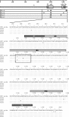
FIG. 1.
FIG. 1.
Sequence analysis of MHV viruses cultured in quadruplicate for 10 passages in the continuous presence of the HR2 entry inhibitor. (Upper) Schematic representation of the MHV-A59 spike protein. The fusion peptide (FP), HR1 and HR2 regions, and transmembrane region (TM) are indicated by gray bars. The furin cleavage site is indicated by an arrowhead. The arrows indicate the positions of the observed mutations in the spike genes of the four viruses (MutA to -D), which were independently passaged in the presence of 10 μM HR2 peptide. (Lower) Sequences of the MHV-A59 spike protein (residues 840 to 1324) with the mutations. Sequences of clones are aligned to the wild-type spike sequence. Dots mark amino acids that are identical. The bars indicate the positions of the FP, HR1, HR2, and TM regions. The boxed sequence marks the 19-residue segment within the HR1 region (residues 988 to 1006) containing 8 out of 14 mutations.
FIG. 2.
FIG. 2.
Effects of HR1 mutations on HR2 peptide susceptibility and virus replication. (A) HR2 peptide susceptibilities of recombinant viruses (RecA to -D) and the parental virus MHV-ERLM (Wt). (B to F) HR2 peptide susceptibilities of mutant viruses (MutA to -D), recombinant viruses (RecA to -D), recombinant viruses containing single-amino-acid substitutions (F988S, A990K, or A1006V), and the parental virus (MHV-ERLM). (G) Single-step growth curve of the parental virus (MHV-ERLM) and recombinant viruses (RecA to -D).
FIG. 3.
FIG. 3.
Ribbon diagram of the fusion core structure of MHV (PDB 1WDF) (20), with the HR1 (residues 969 to 1018) and HR2 (residues 1224 to 1254) peptides in dark and light gray, respectively, and the mutated residues (F988, A990, K997, and A1006) indicated. The insets show modeling of the F988S, A990K, and A1006V substitutions. The magenta arrows point at the green-colored side chains, with faded green representing the substituted side chain (A990K and A1006V) and solid green the original side chain (F988S). The blue arrows indicate the V1226 (lower arrow) and L1228 (upper arrow) side chains, and the red arrow points to the R1237 side chain, with faded green representing the movements of the side chains required to accommodate the V1006-and-K990 side chain.

References

    1. Bosch, B. J., B. E. Martina, R. Van Der Zee, J. Lepault, B. J. Haijema, C. Versluis, A. J. Heck, R. De Groot, A. D. Osterhaus, and P. J. Rottier. 2004. Severe acute respiratory syndrome coronavirus (SARS-CoV) infection inhibition using spike protein heptad repeat-derived peptides. Proc. Natl. Acad. Sci. USA 1018455-8460. - PMC - PubMed
    1. Bosch, B. J., R. van der Zee, C. A. de Haan, and P. J. Rottier. 2003. The coronavirus spike protein is a class I virus fusion protein: structural and functional characterization of the fusion core complex. J. Virol. 778801-8811. - PMC - PubMed
    1. de Haan, C. A., L. van Genne, J. N. Stoop, H. Volders, and P. J. Rottier. 2003. Coronaviruses as vectors: position dependence of foreign gene expression. J. Virol. 7711312-11323. - PMC - PubMed
    1. Duquerroy, S., A. Vigouroux, P. J. Rottier, F. A. Rey, and B. J. Bosch. 2005. Central ions and lateral asparagine/glutamine zippers stabilize the post-fusion hairpin conformation of the SARS coronavirus spike glycoprotein. Virology 335276-285. - PMC - PubMed
    1. Gallo, S. A., K. Sackett, S. S. Rawat, Y. Shai, and R. Blumenthal. 2004. The stability of the intact envelope glycoproteins is a major determinant of sensitivity of HIV/SIV to peptidic fusion inhibitors. J. Mol. Biol. 3409-14. - PubMed

Publication types

LinkOut - more resources