. 2008 Feb;82(4):1851-9.
doi: 10.1128/JVI.02339-07. Epub 2007 Dec 12.

Genome organization and reverse genetic analysis of a type I feline coronavirus

Affiliations

Genome organization and reverse genetic analysis of a type I feline coronavirus

Gergely Tekes et al. J Virol. 2008 Feb.

Abstract

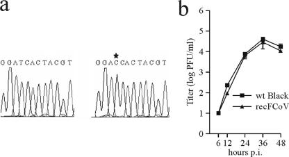
In this study we report the complete sequence and genome organization of the serotype I feline coronavirus (FCoV) strain Black. Furthermore, a reverse genetic system was established for this FCoV strain by cloning a full-length cDNA copy into vaccinia virus. This clone served as basis for the generation of recombinant FCoV (recFCoV) that was shown to bear the same features in vitro as the parental FCoV. Using this system, accessory 3abc genes in the FCoV genome were replaced by green fluorescent protein (recFCoV-GFP) and Renilla luciferase genes (recFCoV-RL). In addition, we showed that feline CD14(+) blood monocytes and dendritic cells can be easily detected after infection with recFCoV-GFP. Thus, our established reverse genetic system provides a suitable tool to study the molecular biology of serotype I FCoV.


Figures

FIG. 1.
FIG. 1.
FCoV type I genome organization. (a) The type I FCoV genome, ORFs, and TRS elements are shown together with the characteristic nested set of viral sgmRNAs. Boxes of viral mRNAs indicate the predicted translationally active “unique” region of each particular mRNA. L, FCoV leader sequence; (A)n, poly(A) tail. (b) Northern blot analysis from poly(A)-containing RNA isolated from type I FCoV strain Black-infected FCWF cells. A 32P-labeled probe directed against the 7ab genes of the type I FCoV genome was used to detect genomic RNA and sgRNAs. (c) Alignment of type I FCoV strain Black TRS elements that direct the synthesis of sgmRNAs. Nucleotides matching the leader TRS are underlined. The conserved TRS core is shaded in gray.
FIG. 2.
FIG. 2.
Schematic diagram for the introduction of the full-length FCoV strain Black cDNA into the vaccinia virus genome. In the first phase, fragments A, D, E, and F (gray boxes) derived from plasmids pA, pD, pE, and pF were ligated into the vaccinia virus (VV) genome (white boxes). In the second phase, to complete the full-length FCoV strain Black cDNA, fragments BC-1, -2, and -3 were introduced using two rounds of vaccinia virus-mediated homologous recombination with gpt-positive and gpt-negative selection using the plasmids pGPT-BC1/3 and pBC-2.
FIG. 3.
FIG. 3.
Analysis of recFCoV. (a) Sequence analysis of the FCoV strain Black and recFCoV in a region containing a silent marker mutation (at nt 485; indicated by an asterisk). (b) Growth kinetics of FCoV strain Black or recFCoV after infection of FCWF cells (MOI of 0.1). wt, wild type; p.i., postinfection.
FIG. 4.
FIG. 4.
Generation and analysis of recFCoV expressing reporter genes. (a) The structural relationship of the FCoV strain Black and the recombinant viruses recFCoV-GFP and recFCoV-RL is shown. ORFs are indicated as boxes. The insertion site of the heterologous reporter gene sequence is depicted together with TRS elements and start and stop codons. (b) Growth kinetics of recFCoV-GFP and recFCoV-RL compared to the parental FCoV strain wt Black (wt) after infection of FCWF cells (MOI of 0.1). (c) Detection of GFP expression in recFCoV-GFP-infected (MOI of 0.1) FCWF cells 24 h postinfection (magnification, ×100; Leica DM IL fluorescence microscope). (d) Kinetics of RL expression in recFCoV-RL-infected (MOI of 0.1) FCWF cells. p.i., postinfection; RLU, relative light units.
FIG. 5.
FIG. 5.
recFCoV-GFP infection of feline monocytic cells. Shown are images of recFCoV-GFP-infected (MOI of 0.1) feline CD14+ monocytes (a) and feline DCs (b) at 24 h postinfection (Leica DM IL fluorescence microscope; magnification, ×100).

References

    1. Addie, D. D., and O. Jarrett. 2001. Use of a reverse-transcriptase polymerase chain reaction for monitoring the shedding of feline coronavirus by healthy cats. Vet. Rec. 148649-653. - PubMed
    1. Addie, D. D., I. A. Schaap, L. Nicolson, and O. Jarrett. 2003. Persistence and transmission of natural type I feline coronavirus infection. J. Gen. Virol. 842735-2744. - PubMed
    1. Berg, A. L., K. Ekman, S. Belak, and M. Berg. 2005. Cellular composition and interferon-gamma expression of the local inflammatory response in feline infectious peritonitis (FIP). Vet. Microbiol. 11115-23. - PMC - PubMed
    1. Black, J. W. 1980. Recovery and in vitro cultivation of a coronavirus from laboratory-induced cases of feline infectious peritonitis (FIP). Vet. Med. Small Anim. Clin. 75811-814. - PubMed
    1. Casais, R., V. Thiel, S. G. Siddell, D. Cavanagh, and P. Britton. 2001. Reverse genetics system for the avian coronavirus infectious bronchitis virus. J. Virol. 7512359-12369. - PMC - PubMed

Publication types