. 2008 Feb;82(4):1665-78.
doi: 10.1128/JVI.02113-07. Epub 2007 Dec 12.

Identification of three interferon-inducible cellular enzymes that inhibit the replication of hepatitis C virus

Affiliations

Identification of three interferon-inducible cellular enzymes that inhibit the replication of hepatitis C virus

Dong Jiang et al. J Virol. 2008 Feb.

Abstract

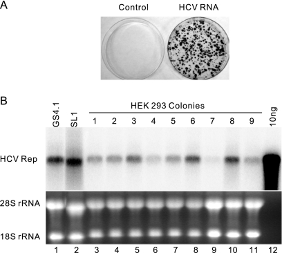
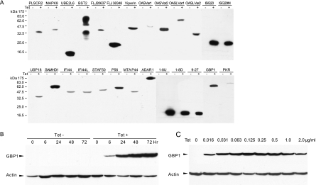
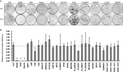
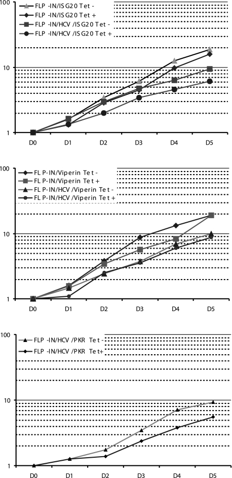
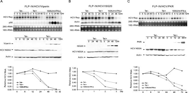
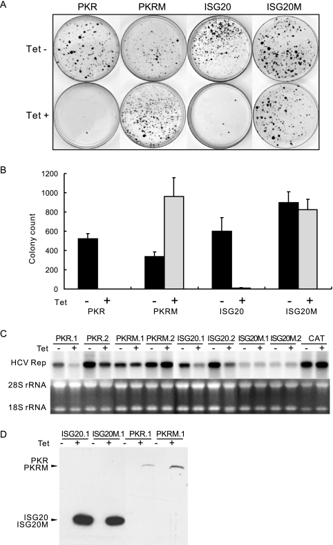
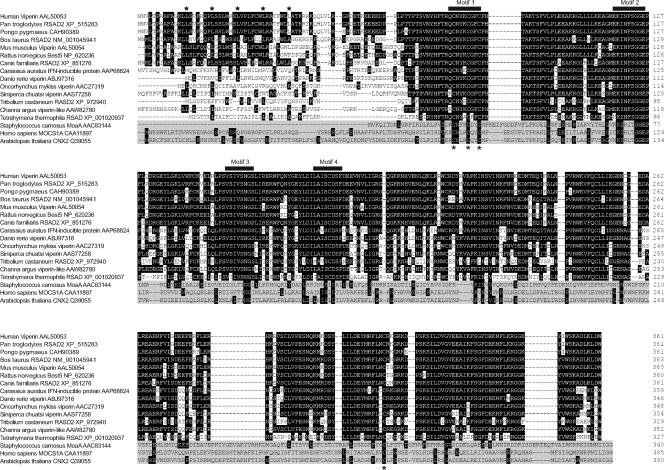
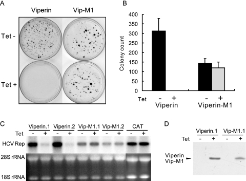
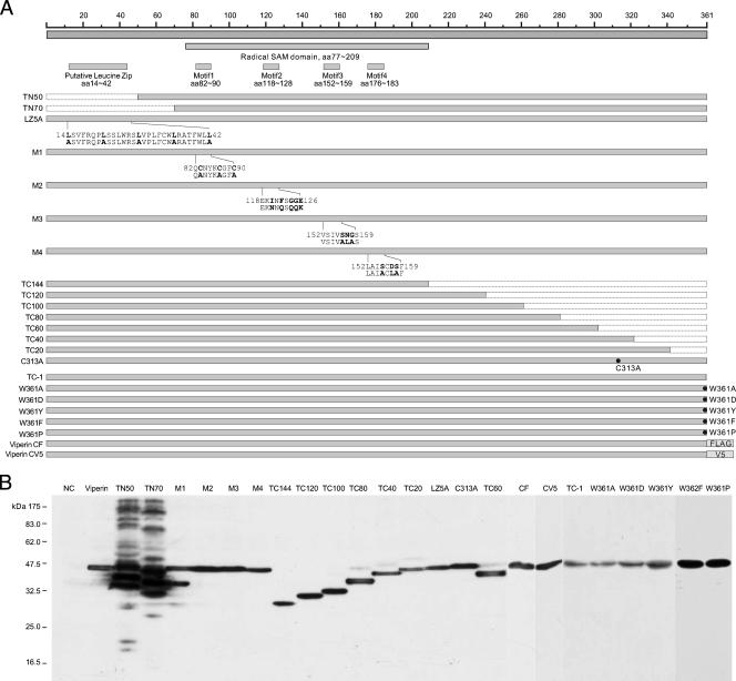
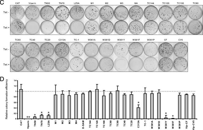
Hepatitis C virus (HCV) infection is a common cause of chronic hepatitis and is currently treated with alpha interferon (IFN-alpha)-based therapies. However, the underlying mechanism of IFN-alpha therapy remains to be elucidated. To identify the cellular proteins that mediate the antiviral effects of IFN-alpha, we created a HEK293-based cell culture system to inducibly express individual interferon-stimulated genes (ISGs) and determined their antiviral effects against HCV. By screening 29 ISGs that are induced in Huh7 cells by IFN-alpha and/or up-regulated in HCV-infected livers, we discovered that viperin, ISG20, and double-stranded RNA-dependent protein kinase (PKR) noncytolytically inhibited the replication of HCV replicons. Mechanistically, inhibition of HCV replication by ISG20 and PKR depends on their 3'-5' exonuclease and protein kinase activities, respectively. Moreover, our work, for the first time, provides strong evidence suggesting that viperin is a putative radical S-adenosyl-l-methionine (SAM) enzyme. In addition to demonstrating that the antiviral activity of viperin depends on its radical SAM domain, which contains conserved motifs to coordinate [4Fe-4S] cluster and cofactor SAM and is essential for its enzymatic activity, mutagenesis studies also revealed that viperin requires an aromatic amino acid residue at its C terminus for proper antiviral function. Furthermore, although the N-terminal 70 amino acid residues of viperin are not absolutely required, deletion of this region significantly compromises its antiviral activity against HCV. Our findings suggest that viperin represents a novel antiviral pathway that works together with other antiviral proteins, such as ISG20 and PKR, to mediate the IFN response against HCV infection.


Figures

FIG. 1.
FIG. 1.
HCV replicons efficiently replicate in FLP-IN T Rex-derived cells. (A) Cell colony formation conferred by SL1 cell-derived HCV replicon replication. FLP-IN T Rex cells were electroporated with total RNA extracted from parental HeLa cells (left) and SL1 cells (right) and selected with medium containing 500 μg/ml of G418 for 3 weeks. Cell foci were stained with crystal violet, and a photograph is shown. (B) GS4.1 and SL1 are Huh7- and HeLa-derived cell lines expressing HCV subgenomic replicons, respectively. Ten micrograms of total RNA isolated from GS4.1, SL1, and nine HEK293HCVrep (lane 3 to 11) cell lines that were established from G418-resistant cell colonies of FLP-IN T Rex cells transfected with SL1-derived RNA was analyzed by Northern blot analysis with an [α-32P]UTP-labeled riboprobe that is complementary to the plus strand of the HCV NS3-coding region. In vitro-transcribed HCV replicon RNA (10 ng; lane 12) served as a molecular weight marker (M) and hybridization control. rRNAs served as loading controls.
FIG. 2.
FIG. 2.
Characterization of ISG expression following tetracycline induction in FLP-IN/ISG cell lines. (A) Establishment of stable cell lines that inducibly express individual ISG proteins. FLP-IN T Rex cell lines that inducibly express individual ISGs were established as described in Materials and Methods. The cells were cultured in the absence or presence of tetracycline for 48 h and then harvested. The levels of N-terminal FLAG-tagged ISG protein expression in cell lysates were determined by Western blot analysis with a monoclonal antibody against FLAG tag. (B) Kinetics of ISG induction. A stable cell line that expresses GBP1 (FLP-IN/GBP1) was cultured in the absence or presence of 1 μg/ml tetracycline, and cells were harvested at the indicated time after the addition of the antibiotic. The levels of GBP1 protein expression in cell lysates were determined by Western blot analysis with a monoclonal antibody against FLAG epitope tag (Sigma). (C) Dose response of tetracycline. FLP-IN/GBP1 cells were cultured in the absence and presence of indicated concentrations of tetracycline for 48 h, and cells were then harvested. The levels of GBP1 protein expression in cell lysates were determined by Western blot analysis. β-Actin served as a loading control and was detected by using a monoclonal antibody against human β-actin.
FIG. 3.
FIG. 3.
Effects of ISG expression on HCV replicon-dependent colony formation. (A) Effects of ISG expression on colony formation. CAT- and ISG-expressing FLP-IN T Rex stable cell lines were electroporated with total RNA extracted from SL1 cells and selected with G418 in the absence or presence of 1 μg/ml tetracycline for 2 to 3 weeks as described in Materials and Methods. Cell foci were stained with crystal violet and photographed. A pair of the representative plates from each of the 10 ISG- and control protein CAT-expressing cell lines that were cultured in the absence (upper panel) or presence (lower panel) of tetracycline is presented. (B) Effects of ISG expression on efficiency of HCV replicating cell colony formation. The numbers of cell foci were counted from three plates cultured in either the absence or presence of tetracycline from each of the ISG- and CAT-expressing cell lines. The RCFE was expressed and plotted as a ratio of the cell foci number obtained from cells that were selected in the presence of tetracycline over that obtained from cells that were cultured in the absence of the antibiotic. *, P < 0.01.
FIG. 4.
FIG. 4.
Effects of viperin, ISG20, and PKR on cell growth. Indicated cell lines were seeded in six-well plates at a density of 1 × 10⁵ cells per well in medium with or without 1 μg/ml tetracycline. Three wells of cells from each of the cell lines cultured with or without tetracycline were trypsinized at 1, 2, 3, 4, and 5 days after seeding. Average rates of cell growth related to cell numbers seeded were plotted.
FIG. 5.
FIG. 5.
Effects of viperin, ISG20, and PKR expression on replication of HCV replicons in HEK293 cells. FLP-IN/HCV/viperin (A), FLP-IN/HCV/ISG20 (B), and FLP-IN/HCV/PKR (C) cell lines were cultured in the absence or presence of 1 μg/ml tetracycline or treated with 100 IU/ml IFN-α in the absence of the antibiotic for 3 days. Cells were harvested at the indicated time points. Ten micrograms of total RNA was analyzed by Northern blot hybridizations with an [α-32P]UTP-labeled riboprobe that is complementary to the plus strand of the HCV NS3-coding region. rRNA and β-actin mRNA served as loading controls (upper panels of A, B, and C). Expression of viperin, ISG20, and PKR and levels of HCV NS5A in the cell lysates were determined by Western blot analyses with antibodies against FLAG tag or HCV NS5A. β-Actin served as a loading control (middle panels of A, B, and C). The amounts of HCV RNA were quantified with the help of a Quantity-One PhosphorImager (Bio-Rad), and the values were plotted as the fraction (percent) of the values obtained with pretreated cells (lower panels of A, B, and C).
FIG. 6.
FIG. 6.
Requirements of the enzymatic activities of PKR and ISG20 on their antiviral effects against HCV. (A) Stable FLP-IN T Rex-derived cells lines that inducibly express N-terminal FLAG-tagged wild-type and enzymatically inactive mutant PKR (PKRM) and ISG20 (ISG20M) were electroporated with total RNA extracted from SL1 cells and selected with G418 in the absence or presence of 1 μg/ml tetracycline for 3 weeks as described in Materials and Methods. Cell foci were stained with crystal violet and photographed. A pair of the representative plates from each of cell lines that were cultured in the absence (upper panel) or presence (lower panel) of tetracycline is presented. (B) Averages and standard derivations of cell foci numbers obtained from three plates of each cell line under the indicated selection conditions were plotted. (C) Two independent HCV replicon-containing cell lines that inducibly express wild-type and mutant PKR or ISG20 were cultured in the absence or presence of 1 μg/ml tetracycline for 3 days. Cells were then harvested, and 10 μg of total RNA was analyzed by Northern blot hybridizations with an [α-32P]UTP-labeled riboprobe that is complementary to the plus strand of the HCV NS3-coding region. rRNA served as a loading control. (D) Inducible expression of wild-type and mutant PKR and ISG20 proteins in the stable cell lines was determined by Western blot analysis with a monoclonal antibody against FLAG tag.
FIG. 7.
FIG. 7.
Multiple alignments of amino acid sequences of viperins from 14 animal species and three representative radical SAM enzymes. The names of the species and enzymes and the GenBank accession numbers of their proteins are indicated on the left. The putative leucine zip and four conserved motifs identified in radical SAM enzymes are highlighted. The sequences of the three radical SAM enzymes are shaded.
FIG. 8.
FIG. 8.
Viperin is a putative radical SAM enzyme. (A) Stable FLP-IN T Rex-derived cell lines that inducibly express N-terminal FLAG-tagged wild-type viperin and the mutant viperin M1 were electroporated with total RNA extracted from SL1 cells and selected with G418 in the absence or presence of 1 μg/ml tetracycline for 3 weeks as described in Materials and Methods. Cell foci were stained with crystal violet and photographed. A pair of the representative plates from each cell line that were cultured in the absence (upper panel) or presence (lower panel) of tetracycline is presented. (B) Averages and standard derivations of cell foci numbers obtained from three plates of each cell line under the indicated selection conditions were plotted. (C) HCV replicon-containing cell lines that inducibly express wild-type and M1 mutant viperin were cultured in the absence or presence of 1 μg/ml tetracycline for 3 days. Cells were then harvested, and 10 μg of total RNA was analyzed by Northern blot hybridizations with an [α-32P]UTP-labeled riboprobe that is complementary to the plus strand of the HCV NS3-coding region. rRNA served as a loading control. (D) Inducible expression of wild-type and M1 mutant viperin in the stable cell lines was determined by Western blot analysis with a monoclonal antibody against FLAG tag.
FIG. 9.
FIG. 9.
Structural and functional analysis of viperin. (A) Schematic representation of the structures of viperin and its mutants. The top panels show the overall structural organization of viperin. The positions of the putative leucine zip motif, radical SAM domain, and four conserved motifs identified in radical SAM enzymes are indicated. The lower panels highlight the nature of the mutations in 23 viperin mutants, which include sequential deletions from the N terminal (TN50 and TN70) and C terminal (TC1 to TC144), point mutations in the four conserved motifs of radical SAM enzymes (M1 to M4), a leucine zip motif (LZ5), and Cys313 (C313A) and Trp361 at the C terminus (W361A, W361D, W361Y, W361F, and W361P). The nature of the point mutations is highlighted in bold underneath the sequences. In addition, two mutant viperins were constructed by adding FLAG or V5 epitope tag at the C terminus (CF and CV5). (B) Inducible expression of mutant viperins in FLP-IN T Rex cells. Cells were transfected with plasmids expressing individual mutant viperins and cultured in the presence of 1 μg/ml of tetracycline for 2 days, and the levels of FLAG-tagged viperin and its mutants in cell lysates were determined by Western blot analysis with a monoclonal antibody against FLAG tag. C-terminal V5-tagged viperin was detected with an antibody against V5 tag. (C) Stable cell lines that inducibly express CAT or wild-type and mutant viperins were electroporated with total RNA extracted from SL1 cells and selected with G418 in the absence or presence of 1 μg/ml tetracycline for 3 weeks as described in Materials and Methods. Cell foci were stained with crystal violet and photographed. A pair of the representative plates from each of 25 cell lines that were cultured in the absence (upper panel) or presence (lower panel) of tetracycline is presented. (D) The numbers of cell foci were counted from three plates from each of the cell lines cultured under both selection conditions. RCFE is defined in the legend of Fig. 3. *, P < 0.05; **, P < 0.01.
FIG. 9.
FIG. 9.
Structural and functional analysis of viperin. (A) Schematic representation of the structures of viperin and its mutants. The top panels show the overall structural organization of viperin. The positions of the putative leucine zip motif, radical SAM domain, and four conserved motifs identified in radical SAM enzymes are indicated. The lower panels highlight the nature of the mutations in 23 viperin mutants, which include sequential deletions from the N terminal (TN50 and TN70) and C terminal (TC1 to TC144), point mutations in the four conserved motifs of radical SAM enzymes (M1 to M4), a leucine zip motif (LZ5), and Cys313 (C313A) and Trp361 at the C terminus (W361A, W361D, W361Y, W361F, and W361P). The nature of the point mutations is highlighted in bold underneath the sequences. In addition, two mutant viperins were constructed by adding FLAG or V5 epitope tag at the C terminus (CF and CV5). (B) Inducible expression of mutant viperins in FLP-IN T Rex cells. Cells were transfected with plasmids expressing individual mutant viperins and cultured in the presence of 1 μg/ml of tetracycline for 2 days, and the levels of FLAG-tagged viperin and its mutants in cell lysates were determined by Western blot analysis with a monoclonal antibody against FLAG tag. C-terminal V5-tagged viperin was detected with an antibody against V5 tag. (C) Stable cell lines that inducibly express CAT or wild-type and mutant viperins were electroporated with total RNA extracted from SL1 cells and selected with G418 in the absence or presence of 1 μg/ml tetracycline for 3 weeks as described in Materials and Methods. Cell foci were stained with crystal violet and photographed. A pair of the representative plates from each of 25 cell lines that were cultured in the absence (upper panel) or presence (lower panel) of tetracycline is presented. (D) The numbers of cell foci were counted from three plates from each of the cell lines cultured under both selection conditions. RCFE is defined in the legend of Fig. 3. *, P < 0.05; **, P < 0.01.

References

    1. Ali, S., C. Pellerin, D. Lamarre, and G. Kukolj. 2004. Hepatitis C virus subgenomic replicons in the human embryonic kidney 293 cell line. J. Virol. 78491-501. - PMC - PubMed
    1. Alter, H. J., and L. B. Seeff. 2000. Recovery, persistence, and sequelae in hepatitis C virus infection: a perspective on long-term outcome. Semin. Liver Dis. 2017-35. - PubMed
    1. Barber, G. N. 2005. The dsRNA-dependent protein kinase, PKR and cell death. Cell Death Differ. 12563-570. - PubMed
    1. Berkovitch, F., Y. Nicolet, J. T. Wan, J. T. Jarrett, and C. L. Drennan. 2004. Crystal structure of biotin synthase, an S-adenosylmethionine-dependent radical enzyme. Science 30376-79. - PMC - PubMed
    1. Bigger, C. B., K. M. Brasky, and R. E. Lanford. 2001. DNA microarray analysis of chimpanzee liver during acute resolving hepatitis C virus infection. J. Virol. 757059-7066. - PMC - PubMed

Publication types

MeSH terms

LinkOut - more resources