Review
. 2008 Aug;49(8):1607-20.
doi: 10.1194/jlr.R700018-JLR200. Epub 2007 Dec 12.

Cardiolipin synthesis for the assembly of bacterial and mitochondrial membranes

Affiliations
Review

Cardiolipin synthesis for the assembly of bacterial and mitochondrial membranes

Michael Schlame. J Lipid Res. 2008 Aug.

Abstract

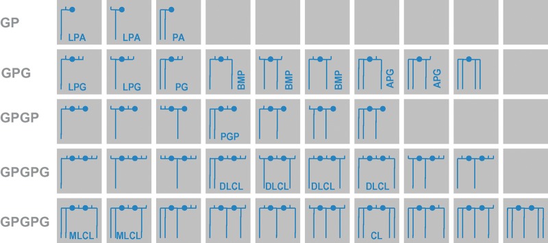
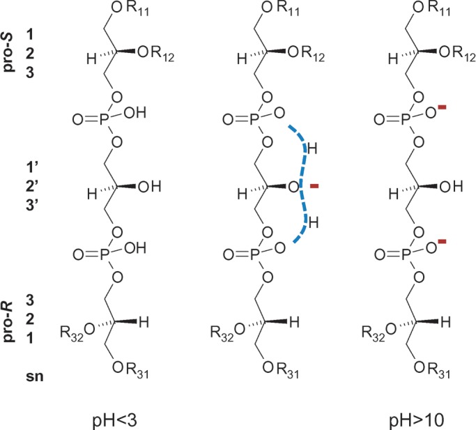
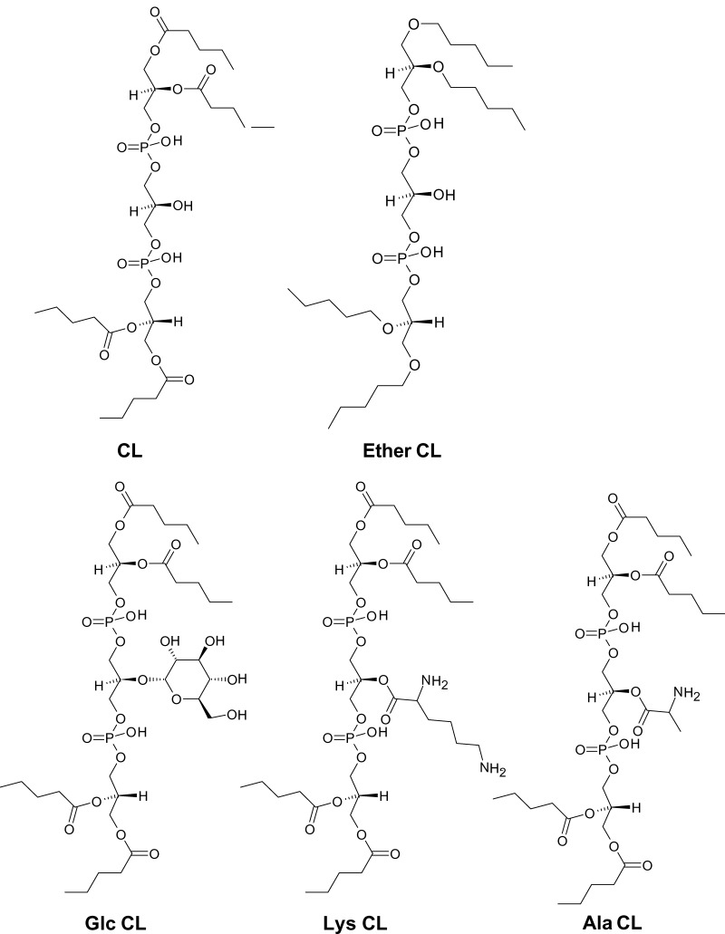
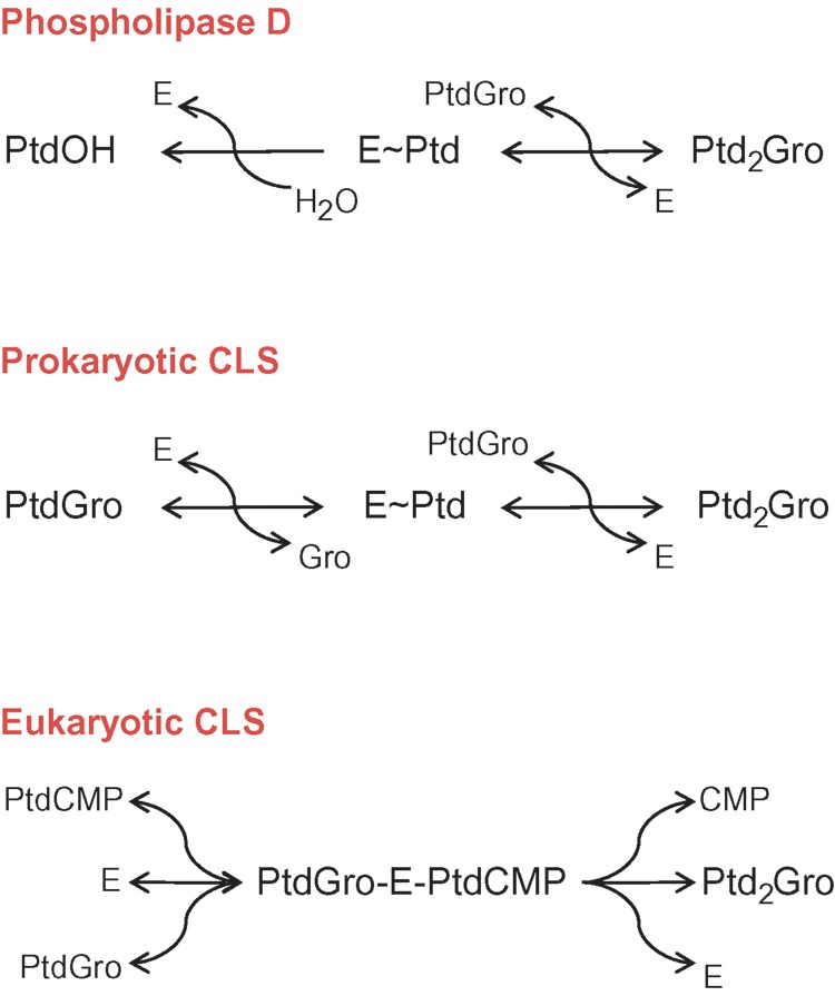
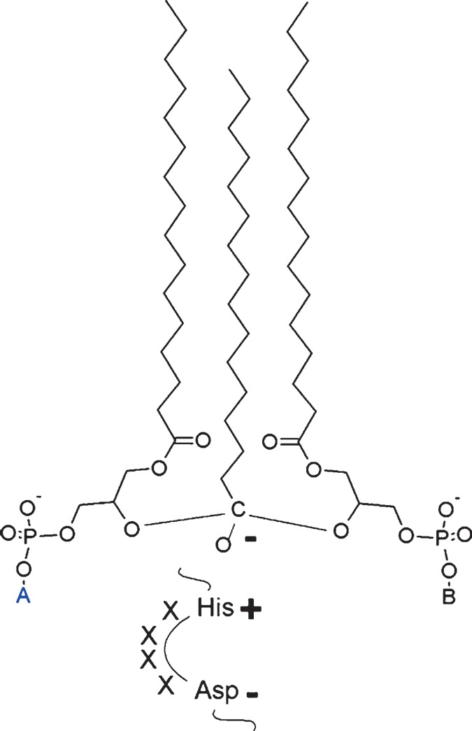
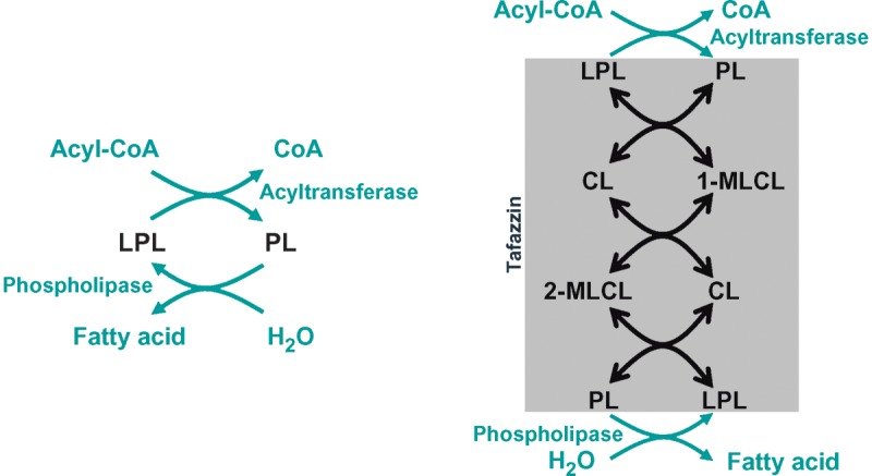
In this article, the formation of prokaryotic and eukaryotic cardiolipin is reviewed in light of its biological function. I begin with a detailed account of the structure of cardiolipin, its stereochemistry, and the resulting physical properties, and I present structural analogs of cardiolipin that occur in some organisms. Then I continue to discuss i) the de novo formation of cardiolipin, ii) its acyl remodeling, iii) the assembly of cardiolipin into biological membranes, and iv) the degradation of cardiolipin, which may be involved in apoptosis and mitochondrial fusion. Thus, this article covers the entire metabolic cycle of this unique phospholipid. It is shown that mitochondria produce cardiolipin species with a high degree of structural uniformity and molecular symmetry, among which there is often a dominant form with four identical acyl chains. The subsequent assembly of cardiolipin into functional membranes is largely unknown, but the analysis of crystal structures of membrane proteins has revealed a first glimpse into the underlying principles of cardiolipin-protein interactions. Disturbances of cardiolipin metabolism are crucial in the pathophysiology of human Barth syndrome and perhaps also play a role in diabetes and ischemic heart disease.


Figures

Fig. 1.
Fig. 1.
Family of polyglycerophospholipids. These lipids consist solely of glycerol groups (horizontal lines), phosphate groups (closed circles), and acyl groups (long vertical lines). Free hydroxyl groups are presented as short vertical lines. The backbones may be glycerophosphate (GP), glycerophospho-glycerol (GPG), glycerophospho-glycerophosphate (GPGP), or glycerophospho-glycero-phosphoglycerol (GPGPG). Stereochemical relationships are not shown in this scheme. The identified compounds have R conformation in the GP group, R/S or S/S conformation in the GPG group, R/S conformation in the GPGP group, and R/R/R, R/S/R, or R/R conformation in the GPGPG group. Members of this family include lysophosphatidic acid (LPA), phosphatidic acid (PA), lysophosphatidylglycerol (LPG), phospatidylglycerol (PG), bis(monoacylglycero)phosphate (BMP), acylphosphatidylglycerol (APG), phospatidylglycerophosphate (PGP), dilysocardiolipin (DLCL), monolysocardiolipin (MLCL), and cardiolipin (CL).
Fig. 2.
Fig. 2.
Structure of cardiolipin. Two phosphatidyl residues, both in R conformation, are linked by a central glycerol bridge. One phosphatidyl group is in pro-R and the other in pro-S position with respect to the central carbon atom. Stereochemical relationships are shown on the left side in sn nomenclature. Different ionic states of the two phosphate groups are presented. R11, R12, R31, and R32 are acyl groups.
Fig. 3.
Fig. 3.
Cardiolipin-like phospholipids from prokaryotes. CL, normal cardiolipin; Ether-CL, tetra-alkylether form of cardiolipin from Halobacterium salinarum; Glc-CL, α-d-glucopyranosyl-cardiolipin from group B Streptococcus; Lys-CL, l-lysyl-cardiolipin from Listeria species; Ala-CL, d-alanyl-cardiolipin from Vagococcus fluvialis.
Fig. 4.
Fig. 4.
Comparison of reaction mechanisms of phospholipase D and cardiolipin synthase. Prokaryotic cardiolipin synthase is a phospholipase D-type enzyme, in which glycerol replaces water as the phosphatidyl acceptor. Note that both steps of prokaryotic cardiolipin synthase are reversible, whereas the hydrolysis step of phospholipase D is irreversible. Eukaryotic cardiolipin synthase is a phosphatidyltransferase that catalyzes an irreversible reaction. E, enzyme; Gro, glycerol; Ptd, 3-sn-phosphatidyl; PtdOH, phosphatidic acid; PtdGro, phosphatidylglycerol; PtdCMP, phosphatidyl-CMP; Ptd2Gro, cardiolipin.
Fig. 5.
Fig. 5.
Sequence alignment of cardiolipin synthases from Saccharomyces cerevisiae, Arabidopsis thaliana, and Homo sapiens. The alignment was computed with CLUSTAL W. Mitochondrial targeting sequences (shown in green) were predicted with MITOPROT (http://ihg.gsf.de). The section corresponding to the general CDP-alcohol phosphotransferase motif is shown in orange. Motifs that are conserved among cardiolipin synthases are shown in red.
Fig. 6.
Fig. 6.
Molecular diversity of cardiolipin species. A total of 81 molecular species can be formed in the presence of three different acyl groups (shown in blue, red, and green). Glycerols are shown in orange, and phosphate groups are shown in black. The diversity arises from the stereochemical nonequivalence of the two phosphatidyl residues in the 1′ and 3′ positions.
Fig. 7.
Fig. 7.
Hypothetical structure of the catalytic intermediate at the active site of tafazzin. In this model, one acyl group is under the simultaneous influence of two lysophospholipid hydroxyl groups. The structure is stabilized by a histidine-aspartate charge relay in accordance with the current model of acyltransferase catalysis. A and B are phospholipid head groups. The catalytic process may progress to form either phospholipid A and lysophospholipid B or lysophospholipid A and phospholipid B.
Fig. 8.
Fig. 8.
Remodeling pathways. Left: Phospholipid remodeling by the deacylation-reacylation cycle. Right: Cardiolipin remodeling by phospholipid transacylation. See text for details. CL, cardiolipin; LPL, lysophospholipid; MLCL, monolysocardiolipin; PL, phospholipid.

References

    1. LeCocq J., and C. E. Ballou. 1964. On the structure of cardiolipin. Biochemistry. 3 976–980. - PubMed
    1. Powell G. L., and J. Jacobus. 1974. The nonequivalence of the phosphorus atoms in cardiolipin. Biochemistry. 13 4024–4026. - PubMed
    1. Schlame M., M. Ren, Y. Xu, M. L. Greenberg, and I. Haller. 2005. Molecular symmetry in mitochondrial cardiolipins. Chem. Phys. Lipids. 138 38–49. - PubMed
    1. Kates M., J-Y. Syz, D. Gosser, and T. H. Haines. 1993. pH-dissociation characteristics of cardiolipin and its 2′-deoxy analogue. Lipids. 28 877–882. - PubMed
    1. Dahlberg M. 2007. Polymorphic phase behavior of cardiolipin derivatives studied by coarse-grained molecular dynamics. J. Phys. Chem. 111 7194–7200. - PubMed

Publication types

LinkOut - more resources