Review
. 2008 May;29(2):219-37.
doi: 10.1016/j.yfrne.2007.08.006. Epub 2007 Oct 15.

Uncovering the mechanisms of estrogen effects on hippocampal function

Affiliations
Review

Uncovering the mechanisms of estrogen effects on hippocampal function

Joanna L Spencer et al. Front Neuroendocrinol. 2008 May.

Abstract

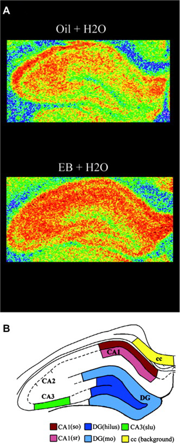
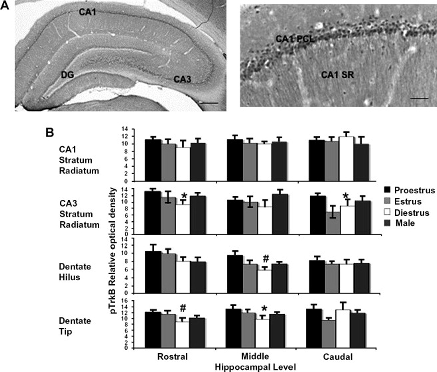
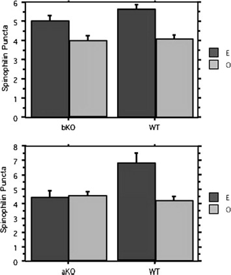
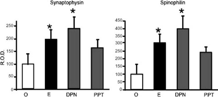
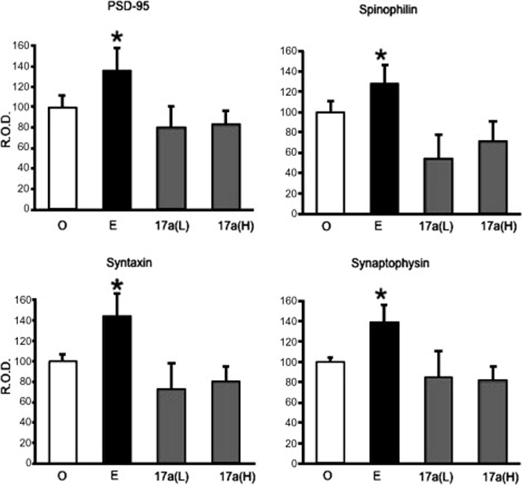
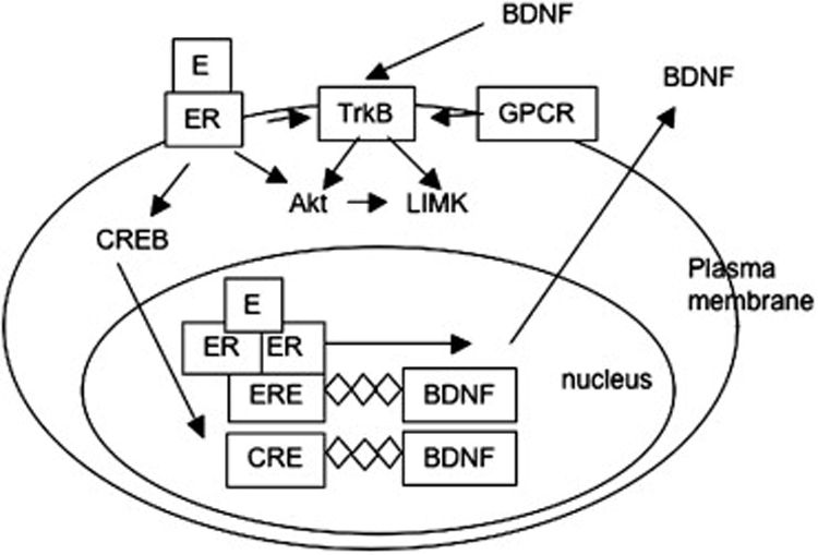
Estrogens have direct effects on the brain areas controlling cognition. One of the most studied of these regions is the dorsal hippocampal formation, which governs the formation of spatial and episodic memories. In laboratory animals, most investigators report that estrogen enhances synaptic plasticity and improves performance on hippocampal-dependent cognitive behaviors. This review summarizes work conducted in our laboratory and others toward identifying estrogen's actions in the hippocampal formation, and the mechanisms for these actions. Physiologic and pharmacologic estrogen affects cognitive behavior in mammals, which may be applicable to human health and disease. The effects of estrogen in the hippocampal formation that lead to modulation of hippocampal function include effects on cell morphology, synapse formation, signaling, and excitability that have been studied in laboratory mice, rats, and primates. Finally, estrogen may signal through both nuclear and extranuclear hippocampal estrogen receptors to achieve its downstream effects.


Figures

Figure 1
Figure 1
Estradiol increases spinophilin expression in the hippocampal formation of ovariectomized female rats. A, Illustration of autoradiograms using pseudocolor representation of shading densities of spinophilin immunoreactivity in the hippocampal formation of representative estrogen (EB)- and control (Oil)-treated ovariectomized rats (blue < green < yellow < orange). B, Schematic diagram identifying specific hippocampal regions from where measures were taken. cc, corpus callosum; dg, dentate gyrus; (so), stratum oriens; (sr), stratum radiatum; (slu); stratum lucidum; (mo), molecular layer. Reprinted with permission from Brake et al., 2001.
Figure 2
Figure 2
pTrkB immunoreactivity (IR) fluctuates across the estrous cycle in the dorsal hippocampal formation of the female rat. A, pTrkB IR in the rat dorsal hippocampal formation. The antibody raised in rabbits against pTrkB was a gift of Moses Chao. IR can be seen in principal cells and processes of the CA1 and CA3 regions of the hippocampus and dentate gyrus (DG). Preadsorption of the antibody with pTrkB blocking peptide eliminates all staining (not shown). B, Matched sections from three different levels of dorsal hippocampus were incubated with rabbit anti-pTrkB at a concentration 1:3000 for 72 hours, labeled by the avidin-biotin complex peroxidase method, and analyzed by densitometry with normalization to corpus callosum staining. Proestrus, n = 7, Estrus, n = 4, Diestrus, n = 4. *p<0.05, #p<0.01 relative to diestrus. Sections correspond to Plates 29 (rostral), 33 (middle), and 36 (caudal) of Paxinos and Watson (Paxinos, G. & Watson, C. The Rat Brain in Stereotaxic Coordinates. Academic Press, San Diego, 1998). Scale Bars, 100 µm. DG, dentate gyrus; PCL, pyramidal cell layer; SR, stratum radiatum.
Figure 3
Figure 3
Estradiol increases spinophilin expression in ovariectomized ER beta knockout mice, but not ER alpha knockout (KO) mice. Ovariectomized female mice were treated with 5 µg estradiol benzoate or oil vehicle for 48 hours and then perfused with 4% paraformaldehyde. Tissue sections stained were for spinophilin using silver-enhanced immunocytochemistry. Data analyzed by two-way Analysis of Variance (ANOVA) showed only an effect of treatment in the ER beta KO mice, with E-treated mice having a higher density of spinophilin immunoreactive puncta than vehicle-treated mice. In the ER alpha KO mice, two-way ANOVA revealed a significant effect of both treatment and genotype, and a significant treatment X genotype interaction. bKO, n = 15 (8 O, 7 E); WT, n = 10 (6 O, 4 E). aKO, n = 11 (6 O, 5 E); WT, n = 7 (3 O, 4 E). O, oil; E, estradiol benzoate; aKO, ER alpha KO; bKO, ER beta KO; WT, wild type; IR, immunoreactive puncta. *p<0.05 relative to O.
Figure 4
Figure 4
ER alpha and beta agonists increase synaptic protein expression in ovariectomized female rats. Ovariectomized female rats were treated for 72 hours with oil vehicle, 10 µg estradiol benzoate, or 5 µg of either PPT (ER alpha agonist) or DPN (ER beta agonist). Tissue lysates from CA1 micropunches were separated by SDS-PAGE and synaptophysin and spinophilin were detected by Western Blotting. The optical density of synaptic protein bands normalized to actin expression was expressed as a percent of the control group. n = 3 animals per group. R.O.D., relative obtical density; EB, estradiol benzoate; O, oil vehicle. *p<0.05 compared to oil-treated group.
Figure 5
Figure 5
17-alpha estradiol does not increase synaptic protein expression in ovariectomized female rats. Rats were treated with 72 hours of 10 µg estradiol benzoate, 15 or 45 µg 17-alpha estradiol, or oil vehicle. Western Blots from punches of hippocampal CA1 area for spinophilin, synaptophysin, syntaxin, and PSD-95 were conducted. Optical density was expressed as a percent of the control (oil) group and normalized to actin expression. O, n = 4; E, n = 4; 17a(L), n = 3; 17a(H), n = 4. O, Oil; E, estradiol benzoate; 17a(L), 17-alpha estradiol, 15 µg; 17a(H), 17-alpha estradiol, 45 µg; R.O.D., relative optical density. *p<0.05 compared to oil.
Figure 6
Figure 6
Estrogen may influence signaling through the TrkB receptor by at least three different mechanisms. Estrogen may induce BDNF transcription by classical action of nuclear hormone receptor through an estrogen response element (ERE) in the BDNF promoter. Alternatively, estrogen actions at extranuclear estrogen receptors (ER) may activate signaling pathways leading to phosphorylation of the CREB protein, leading to transcription through a cAMP response element (CRE) in the BDNF promoter. Finally, estrogen activation at extranuclear ERs may transactivate the TrkB receptor, possibly by coupling to G-protein signaling.

References

    1. Nelson RJ. An Introduction to Behavioral Endocrinology. sunderland, MA: sinauer associates; 2005.
    1. Walf AA, Frye CA. A review and update of mechanisms of estrogen in the hippocampus and amygdala for anxiety and depression behavior. Neuropsychopharmacology. 2006;31:1097–1111. - PMC - PubMed
    1. Korol DL. Role of estrogen in balancing contributions from multiple memory systems. Neurobiol Learn Mem. 2004;82:309–323. - PubMed
    1. Davis DM, Jacobson TK, Aliakbari S, Mizumori SJ. Differential effects of estrogen on hippocampal- and striatal-dependent learning. Neurobiol Learn Mem. 2005;84:132–137. - PubMed
    1. Luine VN, Jacome LF, Maclusky NJ. Rapid enhancement of visual and place memory by estrogens in rats. Endocrinology. 2003;144:2836–2844. - PubMed

Publication types

MeSH terms