Cardiomyocyte cell cycle activation improves cardiac function after myocardial infarction
- PMID: 18079102
- PMCID: PMC2653079
- DOI: 10.1093/cvr/cvm101
Cardiomyocyte cell cycle activation improves cardiac function after myocardial infarction
Abstract
Aims: Cardiomyocyte loss is a major contributor to the decreased cardiac function observed in diseased hearts. Previous studies have shown that cardiomyocyte-restricted cyclin D2 expression resulted in sustained cell cycle activity following myocardial injury in transgenic (MHC-cycD2) mice. Here, we investigated the effects of this cell cycle activation on cardiac function following myocardial infarction (MI).
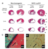
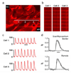
Methods and results: MI was induced in transgenic and non-transgenic mice by left coronary artery occlusion. At 7, 60, and 180 days after MI, left ventricular pressure-volume measurements were recorded and histological analysis was performed. MI had a similar adverse effect on cardiac function in transgenic and non-transgenic mice at 7 days post-injury. No improvement in cardiac function was observed in non-transgenic mice at 60 and 180 days post-MI. In contrast, the transgenic animals exhibited a progressive and marked increase in cardiac function at subsequent time points. Improved cardiac function in the transgenic mice at 60 and 180 days post-MI correlated positively with the presence of newly formed myocardial tissue which was not apparent at 7 days post-MI. Intracellular calcium transient imaging indicated that cardiomyocytes present in the newly formed myocardium participated in a functional syncytium with the remote myocardium.
Conclusion: These findings indicate that cardiomyocyte cell cycle activation leads to improvement of cardiac function and morphology following MI and may represent an important clinical strategy to promote myocardial regeneration.
Conflict of interest statement
Figures


Comment in
-
Divide to survive: myocardial regeneration and functional recovery after cell cycle activation in injured hearts.Cardiovasc Res. 2008 Apr 1;78(1):1-2. doi: 10.1093/cvr/cvn026. Epub 2008 Feb 4. Cardiovasc Res. 2008. PMID: 18250142 No abstract available.
References
-
- Soonpaa MH, Field LJ. Survey of studies examining mammalian cardiomyocyte DNA synthesis. Circ Res. 1998;83:15–26. - PubMed
-
- Oh H, Chi X, Bradfute SB, Mishina Y, Pocius J, Michael LH, et al. Cardiac muscle plasticity in adult and embryo by heart-derived progenitor cells. Ann N Y Acad Sci. 2004;1015:182–189. - PubMed
-
- Anversa P, Nadal-Ginard B. Myocyte renewal and ventricular remodelling. Nature. 2002;415:240–243. - PubMed
-
- Hassink RJ, Brutel de la Riviere A, Mummery CL, Doevendans PA. Transplantation of cells for cardiac repair. J Am Coll Cardiol. 2003;41:711–717. - PubMed
-
- Hassink RJ, Dowell JD, Brutel de la Riviere A, Doevendans PA, Field LJ. Stem cell therapy for ischemic heart disease. Trends Mol Med. 2003;9:436–441. - PubMed
Publication types
MeSH terms
Substances
Grants and funding
LinkOut - more resources
Full Text Sources
Other Literature Sources
Medical
Molecular Biology Databases
Research Materials

