. 2007 Dec 15;21(24):3342-55.
doi: 10.1101/gad.455407.

Mismatch repair-dependent processing of methylation damage gives rise to persistent single-stranded gaps in newly replicated DNA

Affiliations

Mismatch repair-dependent processing of methylation damage gives rise to persistent single-stranded gaps in newly replicated DNA

Nina Mojas et al. Genes Dev. .

Abstract

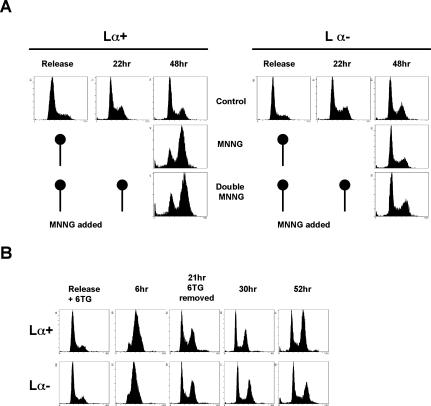
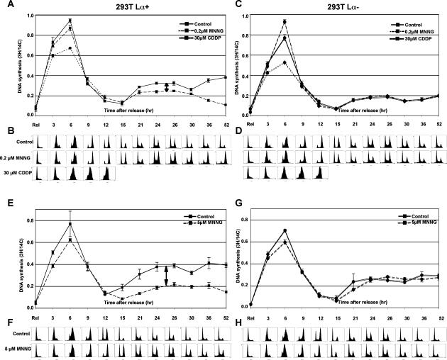
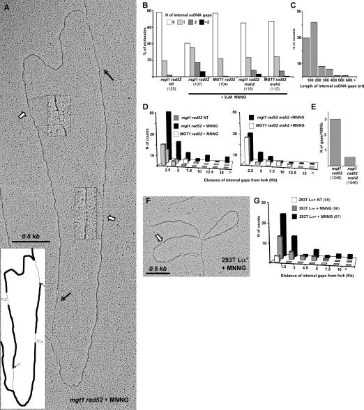
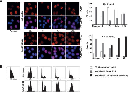
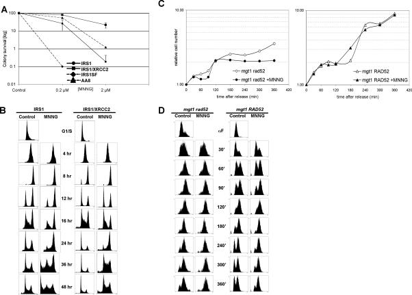
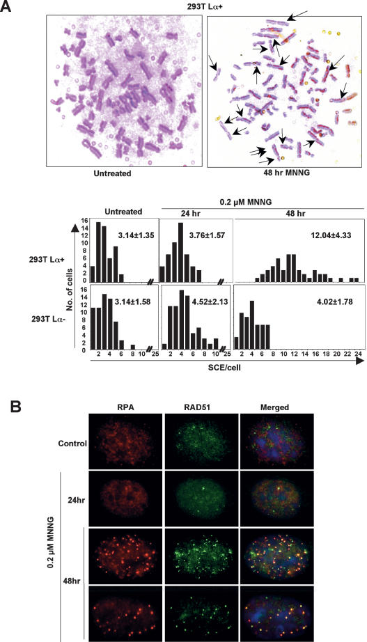
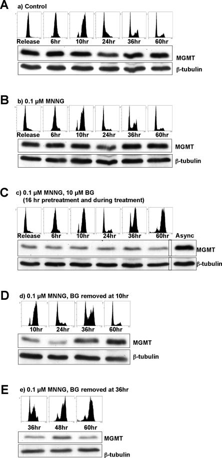
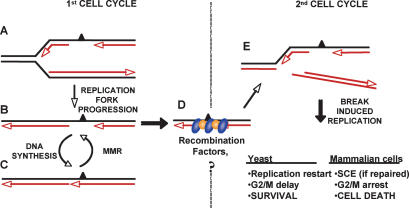
O(6)-Methylguanine ((Me)G) is a highly cytotoxic DNA modification generated by S(N)1-type methylating agents. Despite numerous studies implicating DNA replication, mismatch repair (MMR), and homologous recombination (HR) in (Me)G toxicity, its mode of action has remained elusive. We studied the molecular transactions in the DNA of yeast and mammalian cells treated with N-methyl-N'-nitro-N-nitrosoguanidine (MNNG). Although replication fork progression was unaffected in the first cell cycle after treatment, electron microscopic analysis revealed an accumulation of (Me)G- and MMR-dependent single-stranded DNA (ssDNA) gaps in newly replicated DNA. Progression into the second cell cycle required HR, while the following G(2) arrest required the continued presence of (Me)G. Yeast cells overcame this block, while mammalian cells generally failed to recover, and those that did contained multiple sister chromatid exchanges. Notably, the arrest could be abolished by removal of (Me)G after the first S phase. These new data provide compelling support for the hypothesis that MMR attempts to correct (Me)G/C or (Me)G/T mispairs arising during replication. Due to the persistence of (Me)G in the exposed template strand, repair synthesis cannot take place, which leaves single-stranded gaps behind the replication fork. During the subsequent S phase, these gaps cause replication fork collapse and elicit recombination and cell cycle arrest.


Figures

Figure 1.
Figure 1.
MNNG cell cycle arrest depends on MeG alone and not on other types of methylation damage. (A) Lesions other than MeG do not mediate progression from the second to the third cell cycle. After release from HU synchronization, 293T Lα cells were treated with MNNG and allowed to progress to the following G1 phase, where they were treated with MNNG again in order to create lesions of similar type and quantity as during the first treatment. As can be seen, the presence of these modifications did not influence MMR-dependent arrest, as the cells still arrested in the second G2/M phase after the first treatment. (B) MeTG causes an MMR-dependent arrest identical to that induced by MNNG, suggesting that the cell cycle arrest is activated by methylation of O6 of guanine or S6 of thioguanine, rather than by other types of damage.
Figure 2.
Figure 2.
MMR-dependent processing of MeG does not affect replication fork progression but leads to a reduced replication rate in the second cell cycle. (A,C,E,G) Incorporation rate of tritiated thymidine was measured in 293T Lα cells upon release from HU synchronization after treatment with two different concentrations of MNNG. (B,D,F,H) In parallel, cell cycle progression was followed by DNA content analysis. The incorporation rates during the first cell cycle were similar, indicating that processing of the MeG-containing mispairs by MMR does not affect progression of the replication forks. However, replication efficiency was dramatically affected during the second cell cycle (arrows) in a dose-dependent manner, and only in MMR-proficient cells. All experiments were repeated at least three times in triplicate. The figure shows representative profiles of one single experiment.
Figure 3.
Figure 3.
MMR- and MeG-dependent ssDNA gaps accumulate behind yeast and mammalian replication forks encountering MNNG-damaged templates. (A) Electron micrograph of a representative RI isolated from mgt1 rad52 cells treated with 3 μM MNNG and cross-linked in vivo with psoralen 30 min after release from G1. White arrows and magnified inserts indicate ssDNA regions along the replicated strands (internal gaps), and black arrows indicate the transition from double-stranded DNA (dsDNA) to ssDNA at the replication fork. (B) Distribution of ssDNA gaps in yeast strains treated with 3 μM MNNG. The increased number of gaps was dependent on the presence of MGT1 and functional MMR. The total number of molecules analyzed is shown in parentheses. (C) Lengths of internal gaps in the mgt1 rad52 strain. The majority of the gaps were <200 nt, independently of genetic background (data not shown). (D) Distribution of internal gaps relative to the replication forks. The population of molecules analyzed was the same as in Figure 2B. The increase in the number of gaps far from the fork was dependent on MGT1 and MMR. (E) Number of internal gaps in fully replicated DNA isolated from cells 90 min after release from G1. MMR-proficient cells displayed an increase in the number of gaps along replicated duplexes after MNNG treatment. The total length (in kilobases) of linear replicated DNA analyzed is shown in parentheses. (F) Electron micrograph of a representative RI isolated from 293T Lα+ cells treated with 5 μM MNNG and cross-linked in vivo with psoralen 4 h after release from G1. (G) Distribution of internal gaps relative to replication forks in 293T Lα cells. MNNG treatment brought about an increase in the number of gaps along the replicated duplexes, with 293T Lα+ cells displaying a significant proportion of gaps far from the fork. The total number of molecules analyzed is shown in parentheses. All experiments were carried out at least twice, and the observed differences could be shown to be reproducible. Due to subtle, uncontrollable variations in sample preparation, results from independent experiments cannot be directly averaged. Hence, the figure shows representative graphs from one single experiment.
Figure 4.
Figure 4.
MNNG treatment brings about a retention of PCNA in chromatin. (A) Cells synchronized in G1/S were released and either left untreated or treated with 0.4 μM MNNG. After permeabilization prior to fixation, only chromatin-bound PCNA remained in the cells. (B) Although both control and treated cells progressed through the cell cycle, PCNA was retained in the chromatin only in the MNNG-treated cells. The right panel in A shows a graphic representation of the different types of PCNA staining.
Figure 5.
Figure 5.
Recombination is required for transition from the first into the second cell cycle after MNNG treatment in both mammalian and yeast cells. (A) Colony survival assay of HR-deficient cell lines after MNNG treatment. Cell lines deficient in the RAD51 paralogs XRCC2 (IRS1) and XRCC3 (IRS1SF) are hypersensitive to MNNG compared with their recombination-proficient counterparts. (B) HR-deficient IRS1cells arrested in the first G2 phase after MNNG treatment (12–16 h), whereas the HR-proficient IRS1/XRCC2 cells went through the first cell cycle and arrested in the second G2 after treatment. (C) Growth curves of HR-deficient mgt1 rad52 and HR-proficient mgt1 RAD52 yeast cells. The growth of mgt1 RAD52 cells was unaffected by MNNG treatment, whereas mgt1 rad52 cells stopped growing after one cell cycle. (D) The mgt1 rad52 strain arrests in the first G2/M after MNNG treatment. Although the mgt1 RAD52 cells continued to grow after treatment, they spent longer in G2.
Figure 6.
Figure 6.
Recombination mechanisms in the first and second cell cycles after MNNG treatment differ. (A) Representative images of metaphase spreads of 293T Lα+ cells stained to differentiate sister chromatids of individual chromosomes. Only MMR-proficient cells displayed elevated levels of SCEs, and only 48 h after MNNG treatment. (B) Coimmunostaining of MMR-proficient HeLa cells showing colocalization of RPA and RAD51 foci after 0.2 μM MNNG treatment.
Figure 7.
Figure 7.
MNNG-activated cell cycle arrest requires the persistence of MeG in DNA through two cell cycles. Cells were synchronized with 2 mM HU and released into fresh medium (A) or medium containing 0.1 μM MNNG (B–E). (C) Cells incubated with BG arrested at G2/M two cell cycles after release from HU. (D) When BG was removed 10 h after release (when cells passed the first S phase), the cells continued to proliferate without arresting. (E) In contrast, if BG was removed at the point when the checkpoint was already activated, the cells remained arrested.
Figure 8.
Figure 8.
Model for the roles of replication, MMR, and recombination in DNA transactions induced by MeG. (A) When replication forks encounter MeG in the template DNA of MMR-proficient cells, they insert C or T and proceed with replication. The MeG-containing mispair activates MMR, which degrades the newly synthesized strand up to and some distance past the MeG residue. (B,C) This gap cannot be filled in, because MMR repeatedly inhibits post-replicative gap repair. In the presence of functional recombination, the gaps are protected and progress to the next cell cycle (D), where they cause replication fork collapse (E). The collapsed replication forks can be restored with the help of HR, which leads to cell survival, at a cost of higher SCE levels. Cells that fail to rescue the forks arrest in G2/M and subsequently die because of their inability to restart replication.

References

    1. Berardini M., Mazurek A., Fishel R., Mazurek A., Fishel R., Fishel R. The effect of O6-methylguanine DNA adducts on the adenosine nucleotide switch functions of hMSH2–hMSH6 and hMSH2–hMSH3. J. Biol. Chem. 2000;275:27851–27857. - PubMed
    1. Cejka P., Stojic L., Mojas N., Russell A.M., Heinimann K., Cannavo E., di Pietro M., Marra G., Jiricny J., Stojic L., Mojas N., Russell A.M., Heinimann K., Cannavo E., di Pietro M., Marra G., Jiricny J., Mojas N., Russell A.M., Heinimann K., Cannavo E., di Pietro M., Marra G., Jiricny J., Russell A.M., Heinimann K., Cannavo E., di Pietro M., Marra G., Jiricny J., Heinimann K., Cannavo E., di Pietro M., Marra G., Jiricny J., Cannavo E., di Pietro M., Marra G., Jiricny J., di Pietro M., Marra G., Jiricny J., Marra G., Jiricny J., Jiricny J. Methylation-induced G(2)/M arrest requires a full complement of the mismatch repair protein hMLH1. EMBO J. 2003;22:2245–2254. - PMC - PubMed
    1. Cejka P., Mojas N., Gillet L., Schar P., Jiricny J., Mojas N., Gillet L., Schar P., Jiricny J., Gillet L., Schar P., Jiricny J., Schar P., Jiricny J., Jiricny J. Homologous recombination rescues mismatch-repair-dependent cytotoxicity of S(N)1-type methylating agents in S. cerevisiae. Curr. Biol. 2005;15:1395–1400. - PubMed
    1. Clark A.B., Valle F., Drotschmann K., Gary R.K., Kunkel T.A., Valle F., Drotschmann K., Gary R.K., Kunkel T.A., Drotschmann K., Gary R.K., Kunkel T.A., Gary R.K., Kunkel T.A., Kunkel T.A. Functional interaction of proliferating cell nuclear antigen with MSH2–MSH6 and MSH2–MSH3 complexes. J. Biol. Chem. 2000;275:36498–36501. - PubMed
    1. Dimitrova D.S., Gilbert D.M., Gilbert D.M. Temporally coordinated assembly and disassembly of replication factories in the absence of DNA synthesis. Nat. Cell Biol. 2000;2:686–694. - PMC - PubMed

Publication types