. 2008 Mar;149(3):1407-14.
doi: 10.1210/en.2007-1470. Epub 2007 Dec 13.

Forkhead box protein O1 negatively regulates skeletal myocyte differentiation through degradation of mammalian target of rapamycin pathway components

Affiliations

Forkhead box protein O1 negatively regulates skeletal myocyte differentiation through degradation of mammalian target of rapamycin pathway components

Ai-Luen Wu et al. Endocrinology. 2008 Mar.

Abstract

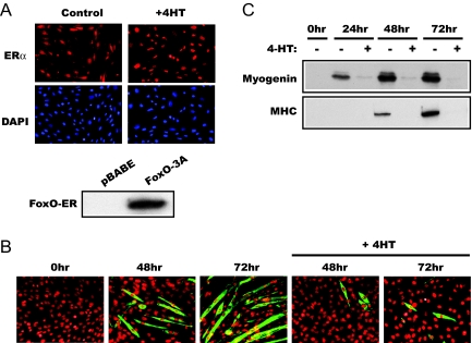
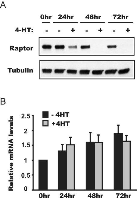
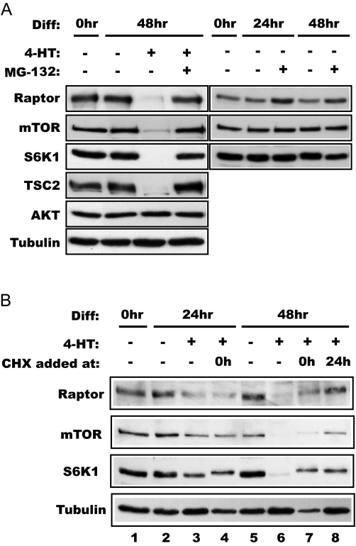
The forkhead transcription factor forkhead box protein O1 (FoxO1), a downstream target of phosphatidylinositol 3-kinase/Akt signaling, has been reported to suppress skeletal myocyte differentiation, but the mechanism by which FoxO1 regulates myogenesis is not fully understood. We have previously demonstrated that a nutrient-sensing mammalian target of rapamycin (mTOR) pathway controls the autocrine production of IGF-II and the subsequent phosphatidylinositol 3-kinase/Akt signaling downstream of IGF-II in myogenesis. Here we report a regulatory loop connecting FoxO1 to the mTOR pathway. Inducible activation of a FoxO1 active mutant in the C2C12 mouse myoblasts blocks myogenic differentiation at an early stage and meanwhile leads to proteasome-dependent degradation of a specific subset of components in the mTOR signaling network, including mTOR, raptor, tuberous sclerosis complex 2, and S6 protein kinase 1. This function of FoxO1 requires new protein synthesis, consistent with the idea that a transcriptional target of FoxO1 may be responsible for the degradation of mTOR. We further show that active FoxO1 inhibits IGF-II expression at the transcriptional activation level, through the modulation of mTOR protein levels. Moreover, the addition of exogenous IGF-II fully rescues myocyte differentiation from FoxO inhibition. Taken together, we propose that the mTOR-IGF-II pathway is a major mediator of FoxO's inhibitory function in skeletal myogenesis.


Figures

Figure 1
Figure 1
Active FoxO1 inhibits C2C12 differentiation. A, C2C12 cells stably expressing FoxO1–3A-ER were immunostained using an anti-ER antibody, accompanied by 4′,6-diamidino-2-phenylindole nuclear staining, before or after 2-h treatment by 1 μm 4-HT. No ER signal was detected in parental C2C12 cells (not shown). The lower panel shows the Western analysis of FoxO1–3A-ER and pBABE cell lysates using the anti-ER antibody. B, FoxO1–3A-ER cells were differentiated in the presence or absence of 4-HT as described in Materials and Methods. At the indicated times, the cells were fixed and immunostained for MHC to reveal myotube formation. C, Lysates of FoxO1–3A-ER cells treated as in B were analyzed by Western blotting for the expression of myogenin and MHC.
Figure 2
Figure 2
Activation of FoxO1 reduces raptor at the protein level. FoxO1–3A-ER cells were differentiated in the presence or absence of 4-HT and lysed at various time points to analyze raptor protein levels by Western blot analysis (A) and mRNA levels by quantitative RT-PCR (B). The average results from three independent experiments are shown, and the error bars represent sd.
Figure 3
Figure 3
Activation of FoxO1 leads to selective loss of proteins in the mTOR signaling pathway. A, FoxO1–3A-ER cells were differentiated in the presence or absence of 4-HT and lysed at various time points for Western blot analyses using the antibodies indicated. B, Cells with the empty vector (pBABE) stably integrated were treated as above and subjected to Western blot analysis. C, C2C12 cells were induced to differentiate in the presence or absence of 50 nm wortmannin (Wort) and lysed at various time points for Western blot analyses using the antibodies indicated. Wortmannin was replenished every 24 h as the differentiation medium was changed.
Figure 4
Figure 4
FoxO1-induced protein degradation is proteasome dependent and requires new protein synthesis. A, FoxO1–3A-ER cells were differentiated (Diff) for 2 d in the presence or absence of 1 μm 4-HT with or without 1 μm MG132 as indicated, followed by Western blot analysis of the lysates. B, The cells were differentiated up to 48 h in the presence of 1 μm 4-HT. Some cells were exposed to 5 μm cycloheximide (CHX) at either 0 or 24 h of differentiation as indicated. The cell lysates were analyzed by Western blotting. Note that the S6K1 band displayed decreased mobility in CHX-treated samples, suggesting phosphorylation of this protein induced by CHX, consistent with reported observations (48).
Figure 5
Figure 5
Activation of FoxO1 suppresses IGF-II expression. A, IGF-II in conditioned media of differentiating FoxO1–3A-ER cells was analyzed by ELISA, and the relative amounts of IGF-II are shown. The average results from three independent experiments are shown, and the error bars represent sd. B, IGF-II ME luciferase reporter was transfected into FoxO1–3A-ER cells, and the luciferase activity was measured at various time points of differentiation in the presence or absence of 4-HT. Data shown are from a representative experiment with triplicate cell treatment for each condition; four independent experiments were performed with similar results.
Figure 6
Figure 6
Exogenous IGF-II rescues myogenic differentiation from inhibition by active FoxO1. A, FoxO1–3A-ER cells were differentiated for 3 d in the presence or absence of 4-HT, with or without 150 ng/ml recombinant IGF-II. Myotube formation was assessed by MHC staining. B, A proposed model: active FoxO suppresses mTOR signaling and subsequent IGF-II production in a feedback loop that modulates myogenesis.

References

    1. Weintraub H 1993 The MyoD family and myogenesis: redundancy, networks, and thresholds. Cell 75:1241–1244 - PubMed
    1. Naya FS, Olson E 1999 MEF2: a transcriptional target for signaling pathways controlling skeletal muscle growth and differentiation. Curr Opin Cell Biol 11:683–688 - PubMed
    1. Florini JR, Ewton DZ, Magri KA 1991 Hormones, growth factors, and myogenic differentiation. Annu Rev Physiol 53:201–216 - PubMed
    1. Barton-Davis ER, Shoturma DI, Musaro A, Rosenthal N, Sweeney HL 1998 Viral mediated expression of insulin-like growth factor I blocks the aging-related loss of skeletal muscle function. Proc Natl Acad Sci USA 95:15603–15607 - PMC - PubMed
    1. Musaro A, McCullagh K, Paul A, Houghton L, Dobrowolny G, Molinaro M, Barton ER, Sweeney HL, Rosenthal N 2001 Localized Igf-1 transgene expression sustains hypertrophy and regeneration in senescent skeletal muscle. Nat Genet 27:195–200 - PubMed

Publication types

MeSH terms