. 2008 Jan;172(1):265-73.
doi: 10.2353/ajpath.2008.070069. Epub 2007 Dec 13.

Nitric oxide regulates neutrophil migration through microparticle formation

Affiliations

Nitric oxide regulates neutrophil migration through microparticle formation

Sarah Nolan et al. Am J Pathol. 2008 Jan.

Abstract

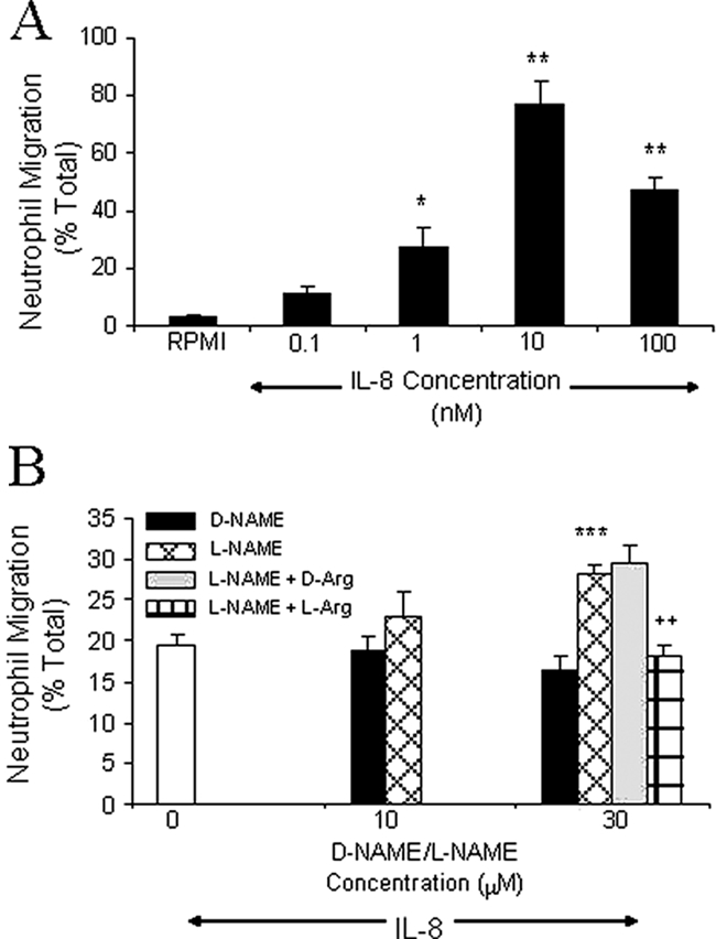
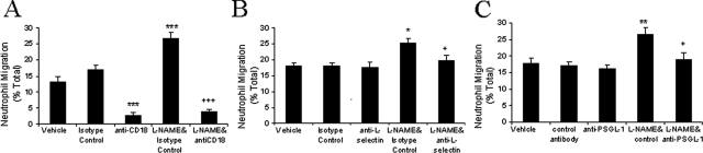
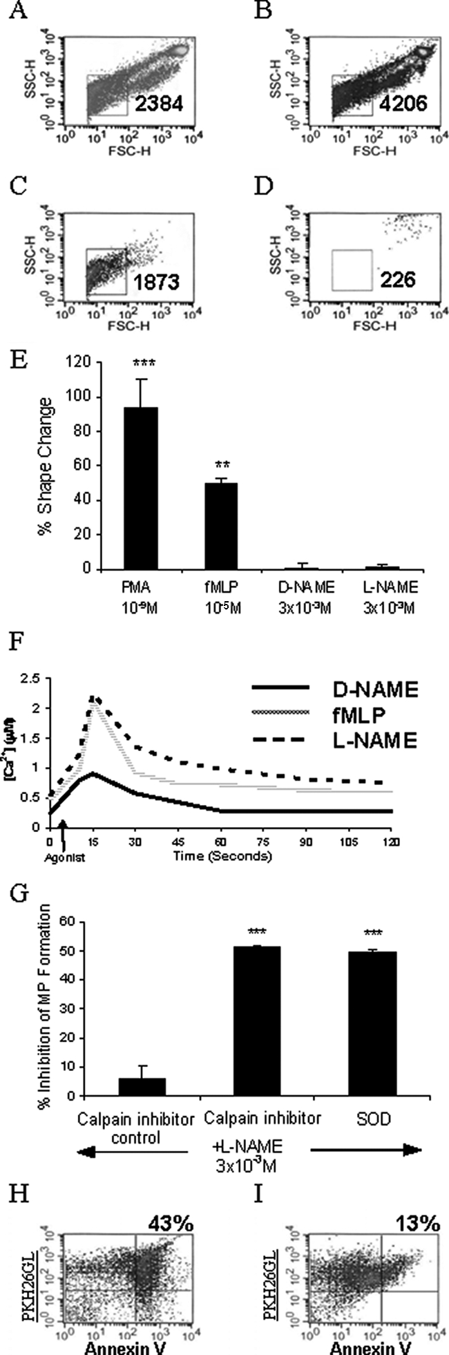
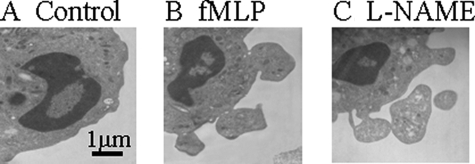
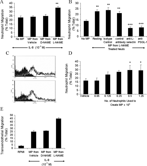
The role of nitric oxide (NO) in regulating neutrophil migration has been investigated. Human neutrophil migration to interleukin (IL)-8 (1 nmol/L) was measured after a 1-hour incubation using a 96-well chemotaxis plate assay. The NO synthase inhibitor N(G)-nitro-l-arginine methyl ester (L-NAME) significantly (P < 0.001) enhanced IL-8-induced migration by up to 45%. Anti-CD18 significantly (P < 0.001) inhibited both IL-8-induced and L-NAME enhanced migration. Antibodies to L-selectin or PSGL-1 had no effect on IL-8-induced migration but prevented the increased migration to IL-8 induced by L-NAME. L-NAME induced generation of neutrophil-derived microparticles that was significantly (P < 0.01) greater than untreated neutrophils or D-NAME. This microparticle formation was dependent on calpain activity and superoxide production. Only microparticles from L-NAME and not untreated or D-NAME-treated neutrophils induced a significant (P < 0.01) increase in IL-8-induced migration and transendothelial migration. Pretreatment of microparticles with antibodies to L-selectin (DREG-200) or PSGL-1 (PL-1) significantly (P < 0.001) inhibited this effect. The ability of L-NAME-induced microparticles to enhance migration was found to be dependent on the number of microparticles produced and not an increase in microparticle surface L-selectin or PSGL-1 expression. These data show that NO can modulate neutrophil migration by regulating microparticle formation.


Figures

Figure 1
Figure 1
Effect of L-NAME on neutrophil migration to IL-8. A: The number of neutrophils that migrated for 1 hour to RPMI or IL-8 (0.1 to 100 nmol/L) was counted and results expressed as a percentage of the total number of neutrophils added to filter membranes of chemotaxis chambers. B: Isolated human neutrophils were resuspended in vehicle (RPMI, open bar), D-NAME, L-NAME, L-NAME with d-arginine (10 mmol/L), or L-NAME with l-arginine (10 mmol/L) and migration to IL-8 (1 nmol/L) assessed after 1 hour. Results are corrected for spontaneous migration by subtracting the percentage of neutrophils migrating to the chemokinesis control. Results are presented as mean ± SEM (n = 3 to 4) and analyzed for statistical significance using one-way analysis of variance followed by Dunnett’s t-test (A) or Bonferroni’s test (B) for multiple comparisons. *P < 0.05, **P < 0.01, and ***P < 0.001 compared to vehicle. ++P < 0.01 compared to the IL-8 response of L-NAME-treated neutrophils.
Figure 2
Figure 2
The effect of adhesion molecule inhibition on L-NAME-induced enhanced migration to IL-8. Neutrophils were resuspended in RPMI, isotype control antibodies, anti-CD18 (6.5E) (A), anti-L-selectin (DREG-200) (B), or blocking and nonblocking anti-PSGL-1 (PL-1, PL-2), with or without L-NAME (C), and added to the filter membrane. Migration to IL-8 was assessed after 1 hour. Data are presented as mean ± SEM (n = 3 to 4) and analyzed for statistical significance using one-way analysis of variance followed by Bonferroni’s test for multiple comparisons. *P < 0.05, **P < 0.01, ***P < 0.001 compared to the IL-8 response of neutrophils treated with isotype control antibody. +P < 0.05, +++P < 0.001 compared to the IL-8 response of neutrophils co-incubated with L-NAME and isotype control.
Figure 3
Figure 3
Analysis of L-NAME-induced microparticle formation. Neutrophils were labeled with PKH26 lipid-intercalating dye (4 μmol/L). Representative flow cytometry density plots showing PKH26-positive events from resting neutrophils (A), neutrophils incubated with L-NAME for 1 hour (B), cell-free supernatant from neutrophils incubated with L-NAME (C), and filtered (0.2 μm) supernatants (D) are shown. Microparticles were identified (square gate) by their smaller forward and side scatter parameters, and events counted for 60 seconds are shown. Density plots are representative of 8 to 13 experiments. E: Gated autofluorescence/forward scatter analysis is shown as mean ± SEM percent change, n = 4. Data were analyzed using one-way analysis of variance followed by Dunnett’s t-test. **P < 0.01, ***P < 0.001 compared to buffer control. F: Flow cytometry analysis of D-NAME, L-NAME, and fMLP effects on intracellular calcium in fluo-3 AM-labeled neutrophils for 2 minutes. Histogram is representative of four experiments. G: Events counted in the microparticle gate for 60 seconds were recorded and the percent inhibition calculated. Results are shown as mean ± SEM, n = 3. Data were analyzed using one-way analysis of variance followed by Dunnett’s t-test. ***P < 0.001 compared to L-NAME-stimulated control. H: Events in the microparticle gate were analyzed for phosphatidylserine externalization by labeling with FITC-conjugated Annexin-V in a calcium-containing buffer. I: As a negative control Annexin-V was added in the presence of EDTA. Microparticles were identified as Annexin V (FL-1)- and PKH26 (FL-2)-positive events located in the top right quadrant on the density plots (percent events shown). The data shown is representative of three experiments.
Figure 4
Figure 4
Electron micrographs showing the formation of microparticles from human neutrophil membranes. Isolated neutrophils were resuspended in RPMI (A), fMLP (B), or L-NAME (C) and incubated for 1 hour. Neutrophils were then fixed in 3% glutaraldehyde-containing buffer and analyzed for microparticle formation.
Figure 5
Figure 5
Microparticles enhance migration to IL-8. Microparticles derived from neutrophils (5 × 10⁶) incubated for 1 hour with RPMI, D-NAME, or L-NAME were resuspended in RPMI (A) or isotype control, nonblocking anti-PSGL-1, anti-L-selectin, or blocking anti-PSGL-1 (B), and migration to IL-8 assessed after 1 hour. C: Microparticle adhesion molecule expression was assessed using PKH26-labeled neutrophils incubated with D-NAME (top) or L-NAME (bottom) for 1 hour. Microparticles were labeled with FITC-anti-L-selectin, FITC-anti-PSGL-1 (shown by the superimposable dark and light gray lines, respectively), or FITC-isotype control (solid area) for 30 minutes at 4°C and analyzed by flow cytometry. Histograms are representative of three experiments. D: The influence of microparticle density was determined by using 0.05 to 1.25 × 10⁶ neutrophils stimulated with L-NAME as a source of microparticles. Migration to IL-8 was measured after 1 hour. Results are presented as mean ± SEM (n = 4). Data were analyzed for statistical significance using one-way analysis of variance followed by Dunnett’s t-test. *P < 0.05, **P < 0.01 compared to the response in the absence of added microparticles, +++P < 0.001 compared to isotype control. E: Migration experiments in A were repeated in the presence of HUVEC monolayer cultured on filter inserts. Results are shown as mean ± SEM from two experiments using two separate blood donors and two separate HUVEC preparations. Each experiment contained four replicates of each treatment.

References

    1. Smith JA. Neutrophils, host defense, and inflammation: a double-edged sword. J Leukoc Biol. 1994;56:672–686. - PubMed
    1. Lukacs NW, Ward PA. Inflammatory mediators, cytokines, and adhesion molecules in pulmonary inflammation and injury. Adv Immunol. 1996;62:257–304. - PubMed
    1. Malik AB, Lo SK. Vascular endothelial adhesion molecules and tissue inflammation. Pharmacol Rev. 1996;48:213–229. - PubMed
    1. Simon SI, Green CE. Molecular mechanics and dynamics of leukocyte recruitment during inflammation. Annu Rev Biomed Eng. 2005;7:151–185. - PubMed
    1. Kakkar AK, Lefer DJ. Leukocyte and endothelial adhesion molecule studies in knockout mice. Curr Opin Pharmacol. 2004;4:154–158. - PubMed

Publication types